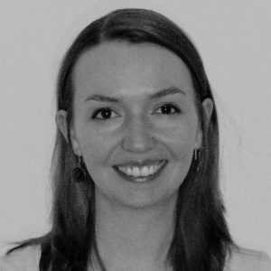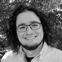
CIBM’s fifth Breakfast & Science Seminar, held on May 26 featured two talks highlighting CIBM researchers’ latest research findings.
Ileana Jelescu, Research Staff Scientist & 14T MRI Operational Manager, CIBM MRI EPFL Animal Imaging and Technology Section presented on “The MRI scanner as a sharp in vivo microscope: from white to gray matter” and Pol del Aguila Pla, Research Staff Scientist, CIBM SP EPFL Mathematical Imaging Section on “GlobalBioIm — Imaging as an inverse problem made easy”. The abstracts are described below, as well as links to recordings of the presentations:
 The MRI scanner as a sharp in vivo microscope: from white to gray matter
Ileana Jelescu,
Research Staff Scientist & 14T MRI Operational Manager, CIBM MRI EPFL Animal Imaging and Technology Section
Abstract:Can we obtain sub-millimeter information about tissue structure vivo and non-invasively? In a diffusion MRI experiment, the signal in encodes information about length scales of a few microns – much smaller than the actual MR image resolution of millimeters – and thus about features of the underlying tissue microstructure that lie in the mesoscale and that we otherwise cannot spatially resolve in vivo. The main challenge of the field is to decode this information and output specific and reliable biophysical parameters of tissue microstructure. In this talk, I will summarize the long and winding road to establishing a reliable biophysical model for diffusion in white matter and illustrate its tremendous potential in a few applications. I will then present our first steps into the next challenge for our field: developing reliable model(s) for cortical and deep gray matter.
 GlobalBioIm. Imaging as an inverse problem made easy
Pol del Aguila Pla,
Research Staff Scientist CIBM SP EPFL, Mathematical Imaging Section
Abstract:Most image reconstruction methods across biomedical imaging rely on the successful formalism of imaging as an inverse problem. In this framework, an imaging system is represented as a transformation of a continuous quantity (e.g., a density of fluorophores over a certain plane or volume) into a number of measurements. Image reconstruction is then understood as the recovery of the original continuous quantity. The successful design of reconstruction methods following this paradigm involves both advanced mathematics to reliably incorporate prior knowledge and major efforts to develop efficient algorithms. In this talk, I explain the basic concepts of the field and show how our library “GlobalBioIm” can facilitate your research, with a couple of running examples to showcase its different functionalities.
The virtual event was well attended, with 57 participants.
In addition to the two interesting presentations, the Breakfast and Science Seminar continued with a ‘round robin’ session where CIBM staff shared insights into each of their sites, which have been difficult to access due to restrictions in place during the containment measure for COVID-19. Staff reported that despite challenges during the worst of the crisis, there has been a recent and gradual return to support the equipment sites.
The latest rules and regulations due to COVID-19 on using CIBM infrastructure are available here:
CIBM MRI CHUV-UNIL (PDF file)
CIBM PET HUG-UNIGE (PDF file)
Towards the end of the Seminar, CIBM MRI CHUV-UNIL Research Staff Scientist , Eleanora Fornari, informed attendees that a new online booking calendar for CIBM CHUV infrastructure is soon to be launched, CIBM Executive Director, Dr. Pina Marziliano confirmed the news and that more details will follow.
Take a look to the upcoming dates for the Breakfast & Science seminars


