Research teams from three of CIBM’s founding institutions have been awarded CHF 3 million to develop the next generation of Positron emission tomography (PET).
Positron emission tomography (PET) is a modern imaging technique that follows radioactive molecules within the human body. Thereby, PET scanners detect small tumors, characterize brain function, and quantify heart perfusion. Now, researchers from the University of Geneva (UNIGE), Écôle polytechnique fédérale de Lausanne (EPFL) and Geneva University Hospitals (HUG) aim to shape the future of PET by introducing a revolutionary scanner design. The SNSF is convinced of their vision for the second time, and supports their project with over CHF 3 million through the SINERGIA scheme that promotes breakthrough research. (www.snf.ch/en/funding/programmes/sinergia).
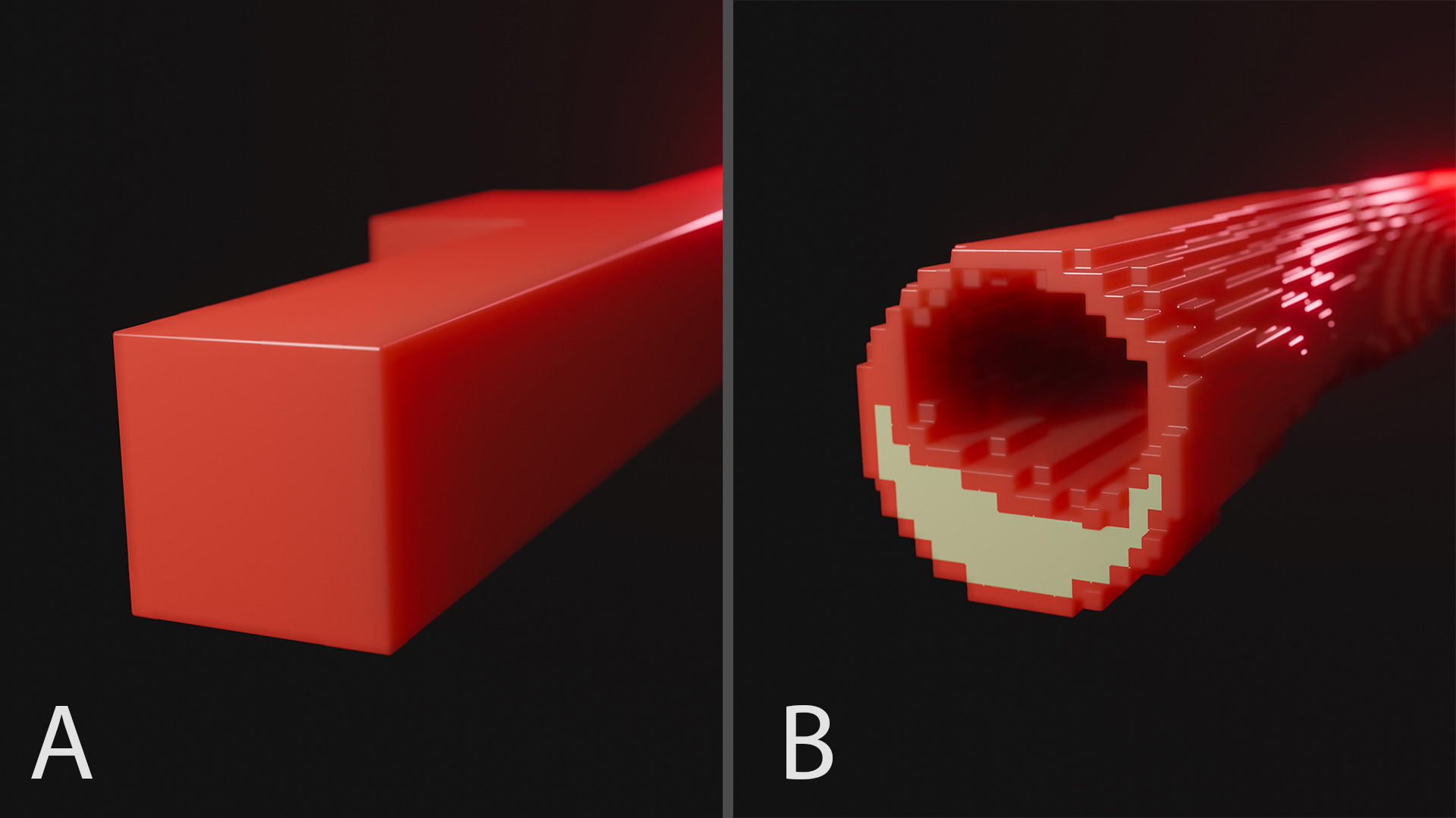
The previously awarded SNSF SINERGIA grant allowed the development of a new type of PET detector, a sensor technology combined with a simulation and image reconstruction framework. The potential of this novel concept is the central part of the new PET scanner.
“We are delighted about the continuous support from the SNSF”, says Prof. Giuseppe Iacobucci (UNIGE, Département de Physique Nucléaire et Corpusculaire and President of the Section of Physics), “that allowed the development of new detectors and the creation of a group of excellent young physicists and engineers. The sensors that we are developing at UNIGE will have many applications in basic science and in the medical domain.” The new PET detector technology is anticipated, for the first time, to explore structures of a few hundred microns, roughly corresponding to the width of a hair .
“The leap in the detector hardware,” adds Prof. Michael Unser (EPFL, Biomedical Imaging Group and Section Head of CIBM Signal Processing EPFL Mathematical Imaging), “challenges us to produce an equal leap in the signal processing software. For the first time, we are using PET to explore objects of such small size, and we need to develop new techniques for the acquisition, reconstruction and understanding of these images.”
The first three years of the project are devoted to developing the prototype of a PET scanner that will be installed at HUG. “The unprecedented accuracy represents a fantastic opportunity that cannot be overestimated,” states Prof. Martin A. Walter (HUG and Section Head of CIBM PET HUG-UNIGE Molecular Imaging). “Our prototype will be used for basic research. As a first application, we aim to generate the first functional PET images of atherosclerotic plaques in mice, which will facilitate a variety of research projects to improve the management of the disease (Figure 1). Our general aim, however, is that every researcher using animal models to improve disease detection, understanding, and treatment will benefit from the new opportunity.”
According to Prof. Pina Marziliano, Executive Director of the CIBM Center for Biomedical Imaging, “The funding of this project demonstrates the outcome of the collaborative efforts between the Core and Affiliate members of CIBM’s founding partner institutions. The multi-disciplinary and translational nature of the project will provide opportunities for developing innovative imaging methods and cutting-edge technologies which will be clinically useful and is aligned with a major CIBM strategic objective.”
To specify the project and write a strong proposal the members of the three teams had many regular zoom meetings. Actively participating and contributing were Dr. Pol del Aguila, CIBM SP EPFL Research Staff Scientist, Dr. Lorenzo Paolozzi, UNIGE Maître Assistant and Olivia Bejuy, CIBM PET HUG-UNIGE Research Staff Scientist who contributed to describe animal models of human diseases that are not imageable with current non-invasive imaging and will highly benefit from high resolution PET in the understanding and treatment of these diseases.
The achievement is aligned with another important CIBM strategic objective, namely, to create opportunities and promote cross-fertilization and multidisciplinarity within the CIBM community, thus allowing younger researchers to grow and be the leaders of tomorrow.