Have you ever imagined cycling inside an MRI scanner while researchers peer into your heart in real-time? The CIBM MRI CHUV-UNIL is excited to announce that this vision has become a reality, thanks to the arrival of a cutting-edge MR-compatible cycling ergometer. This unique setup allows exercise stress tests to be performed directly within the scanner. With its 80 cm-wide bore, the 0.55T low-field MRI scanner accommodates individuals of all heights, enhancing participant comfort and opening exciting new avenues for cardiovascular research.
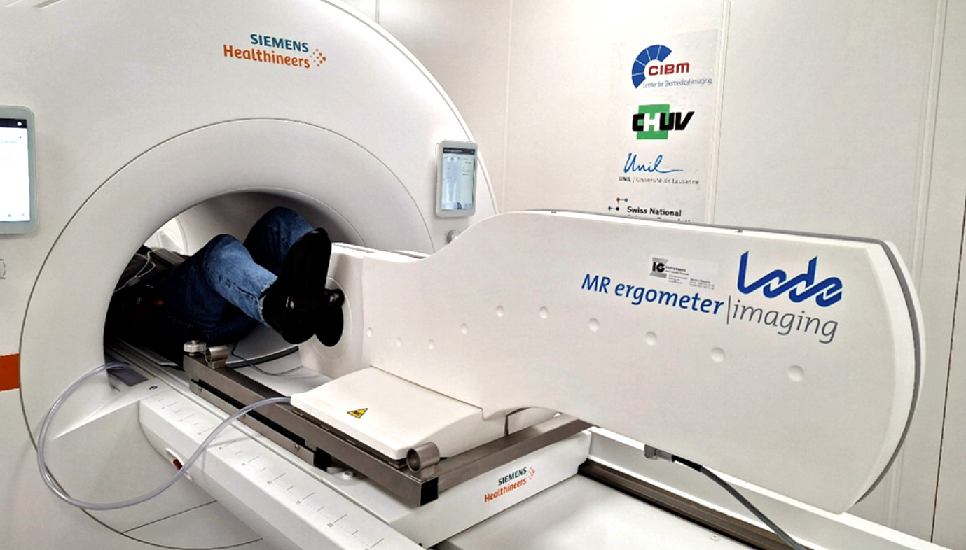
Figure 1: A participant performing exercise stress testing inside the 80 cm-wide bore of the low-field MRI scanner, demonstrating the unique integration of physical activity and real-time imaging. The 80cm-wide bore enables continuous exercise MRI for all subjects, without limitations on subjects’ height
Exercise stress testing is a cornerstone of cardiovascular diagnosis, providing unparalleled insights into the physiology and adaptability of the cardiovascular system. However, combining exercise with MRI in real time has historically been challenging due to the confined space of traditional MRI scanners and the technical complexity of imaging a moving body. Our novel low-field MRI Scanner, operating at 0.55T and equipped with an extra-wide bore, overcomes these obstacles, creating a platform for seamless, in situ plug-and-play exercise MRI. This integration supports real-time imaging during sustainable, controlled, and reversible exercise stress. When paired with emerging faster and motion-compensated MRI technologies, spearheaded by the CIBM research teams, this setup will be the first platform for highly comprehensive exploration of specific cardio-physiological conditions.
The versatility of this system opens new avenues for exploring cardiovascular health and adaptive physiology. From probing organ function to uncovering pathophysiological mechanisms, exercise MRI provides a comprehensive tool to assess the interplay between different physiological systems during a rest-stress protocol, with the ergometer supporting resistance levels up to 300 Watts.
This state-of-the-art infrastructure is a technical milestone and a collaborative resource. We invite researchers from diverse fields to take advantage of this platform for their studies. Whether you are investigating novel biomarkers, validating emerging imaging techniques, or studying specific patient populations, this facility is ready to support your research endeavours.
To get involved, visit our website (Human MRI Research Project Application Form – CIBM | Center for Biomedical Imaging) and submit your research proposals. Let’s pedal into new territories of biomedical research together, advancing our understanding of cardiovascular health and beyond!
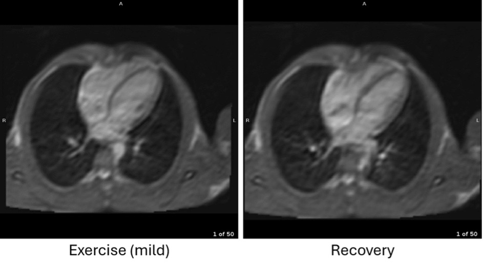
Figure 2 : MRI images of the heart captured using a real-time cardiac MRI protocol during mild exercise (left) and during recovery (right), showcasing the scanner’s ability to visualize cardiac function and anatomy under various physiological conditions.