CIBM SP CHUV-UNIL SECTION

Trustworthy Medical Image Analysis

Section Head: Dr. Meritxell Bach Cuadra, MER, PD (UNIL)

The CIBM Signal Processing CHUV-UNIL Trustworthy Medical Image Analysis Section research centers on the development of advanced image processing and machine learning techniques for medical image computing. We address challenges such as image quality assessment, super-resolution, registration, detection, segmentation, normative modelling, with direct applications in computer-aided diagnosis and therapy planning. The group maintains a strong emphasis on translational research through collaborations with clinical partners in radiology, neurology, oncology, neuroscience, and psychiatry. To support clinical integration, our methods are developed in alignment with emerging regulatory requirements for trustworthy and explainable AI in healthcare. Our section focuses on uncertainty quantification and robust domain adaptation to ensure that AI systems deliver reliable, reproducible results across diverse clinical settings. In parallel, we emphasize human-centered AI design—promoting interpretability and effective communication of model outputs to support clinical decision-making and foster clinician trust.”

RESEARCH TOPICS

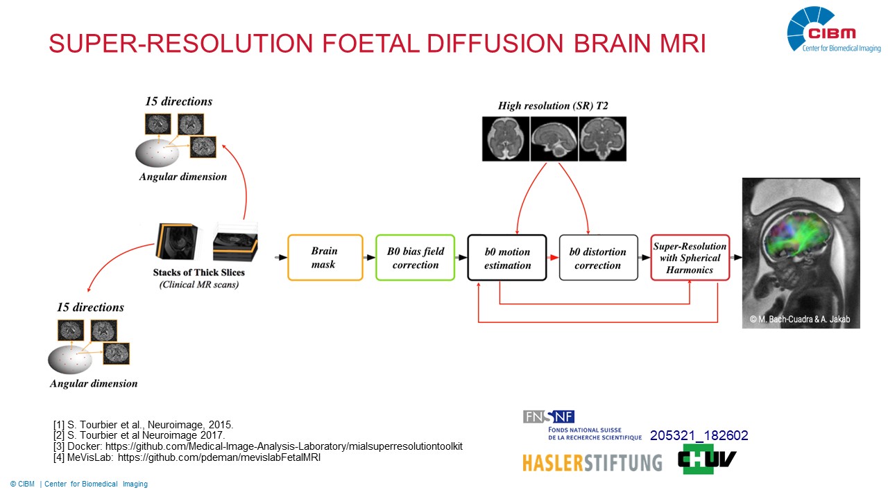
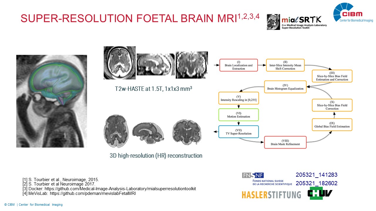
Super-Resolution T2w Foetal Brain MRI for Computer Assisted Diagnosis and Quantitative Analysis

Description: We develop advanced quantitative imaging techniques for studying the maturation of the in-vivo human brain in its early stages of development, when it undergoes the most significant changes. One research line is dedicated to the image super-resolution reconstruction and segmentation of T2w fetal imaging. This will provide accurate quantitative analysis of the growing brain anatomy by morphological biomarkers. Another research line is related to support the translation of the developed reconstruction and segmentation methods to the clinical environment and their evaluation in daily exploration of fetal brain MR images. This research is supported by Swiss National Science Foundation (205321_182602 and 205321_141283), Haslerstiftung and the Radiology Department of the Lausanne University Hospital.

Investigators: Priscille de Dumast (UNIL), Hamza Kebiri (UNIL), Hélène Lajous (UNIL), Meritxell Bach Cuadra (UNIL)

Collaborators: Dr. S. Tourbier (Radiology Department CHUV), Prof. P. Hagmann (Radiology Department CHUV), Prof. P. Maeder (Radiology Department CHUV), Dr V. Dunet (Radiology Department CHUV), Dr. M. Koob (Radiology Department CHUV), Prof. R. Meuli (Radiology Department CHUV), T. Yu (EPFL), Prof. J.-Ph. Thiran (EPFL), Dr. B. Marechal (Siemens Healthcare), M. R. Corredor (Siemens Healthcare)

Super-resolution reconstruction of in-vivo diffusion brain MRI

Description: The assessment of in-vivo fetal brain maturation is not only linked to morphology but also to physiological processes, thus the underlying microstructural changes need also to be imaged. Diffusion MRI (dMRI) allows to depict the underlying tissue microstructure but it suffers from a coarse spatial-resolution. A major research topic is to develop a super-resolution framework encompassing image artefact correction, motion estimation and dMRI reconstruction.  Our work will include spherical harmonics (SH) representation of the signal with spatial-angular regularization and single-volume SR strategies with machine learning-based algorithms. This research is supported by Swiss National Science Foundation (205321_182602 and 205321_141283).

Investigators: Hamza Kebiri (UNIL), Priscille de Dumast (UNIL), Hélène Lajous (UNIL), Meritxell Bach Cuadra (UNIL)

Collaborators: Dr. Y. Alemán-Gómez (Radiology Department CHUV), Dr. S. Tourbier (Radiology Department CHUV), Prof. P. Hagmann (Radiology Department CHUV), Prof. P. Maeder (Radiology Department CHUV), Dr V. Dunet (Radiology Department CHUV), Dr. M. Koob (Radiology Department CHUV), Prof. R. Meuli (Radiology Department CHUV), T. Yu (EPFL), Prof. J.Ph. Thiran (EPFL), Dr. A. Jakab (Kinderspital Zürich)

Quantitative MR Imaging (qMRI) of the in vivo fetal brain

Description: In combination with the planned advancement of foetal T2w and dMR imaging described above, we aim at developing new qMRI sequences for T1 and T2 mapping of the foetal brain. QMRI has proven its ability not only to depict brain maturation but also abnormal patterns and developmental schemes in preterm and new-born babies. The possibility to image these quantitative characteristics in utero would be a major breakthrough in foetal MRI. In this research project, we will combine our SR framework with fast qMRI acquisition schemes tailored to deal with foetal motion. This research is supported by Swiss National Science Foundation (205321_182602 and 205321_141283).

Investigators: Hélène Lajous (UNIL), Hamza Kebiri (UNIL), Priscille de Dumast (UNIL), Hélène Lajous (UNIL), Jean Batiste Ledoux (CHUV), Christopher W. Roy (CHUV), Matthias Stuber (CHUV), Ruud Van Heeswijk (CHUV), Meritxell Bach Cuadra (UNIL)

Collaborators: Dr. S. Tourbier (Radiology Department CHUV), Prof. P. Hagmann (Radiology Department CHUV), Prof. R. Meuli (Radiology Department CHUV), T. Yu (EPFL), Prof. J.-Ph. Thiran (EPFL), Dr. T. Hilbert (Siemens Healthcare), Dr. D. Piccini (Siemens Healthcare), Dr. T. Kober (Siemens Healthcare)

Machine learning methods for MRI analysis in Multiple Sclerosis

Description: We have developed jointly with our collaborators many machine learning methods for the detection and segmentation of white matter and cortical lesions based on MRI of Multiple Sclerosis. Our major contribution has been the development of a partial-volume method to detect small lesions, even in the earliest stages of the disease. Our methods have been developed for clinical MRI setting and also for more advanced MRI sequences at 3T and 7T. Furthermore, we also investigate support diagnosis and prognostic tools based on advanced MRI-based biomarkers (central vein sign and rim sign). This project is supported by European Union’s Horizon 2020 research and innovation program under the Marie Sklodowska-Curie project TRABIT (agreement No 765148).

Investigators:  Francesco La Rosa (EPFL), Hamza Kebiri (UNIL), Meritxell Bach Cuadra (UNIL)

Collaborators: Prof. C. Granziera (University Hospital Basel and University of Basel), Dr. M.J. Fartaria (Siemens Healthcare), Dr J. Richiardi (Siemens Healthcare), Dr. T. Kober (Siemens Healthcare), Dr. P. Maggi (CHUV), Prof. Du Pasquier (CHUV), Prof. R. Meuli (CHUV), Dr. A. Abdulkadir (University of Pennsylvania), Dr. M. Absinta (Johns Hopkins University), Dr E. Beck (Translational Neuroradiology Section – NIH), Dr. P. Sati (Translational Neuroradiology Section – NIH), Dr. D. Reich (Translational Neuroradiology Section – NIH)