CIBM MRI CHUV-UNIL SECTION:

Translational MR Imaging

Section Head: Prof. Matthias Stuber (UNIL)
The CIBM MRI CHUV-UNIL Translational MR Imaging Section includes 17 engineers and physicists who are directly located at the CHUV.  The research effort of this CIBM Section addresses significant human health concerns. It is focused on the development and translational application of novel MRI methodology in strong interdisciplinary collaboration with medical professionals locally, regionally, and internationally. Dissemination of such methodology is one of the declared aims of the MR activity at CHUV-UNIL.

RESEARCH TOPICS

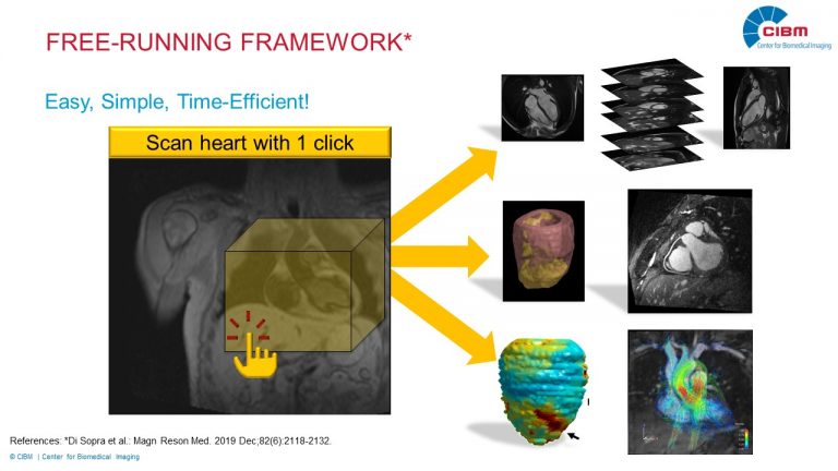
A Paradigm Shift in Magnetic Resonance Imaging of the Heart: 5D Imaging - Sample Now and Ask Questions Later

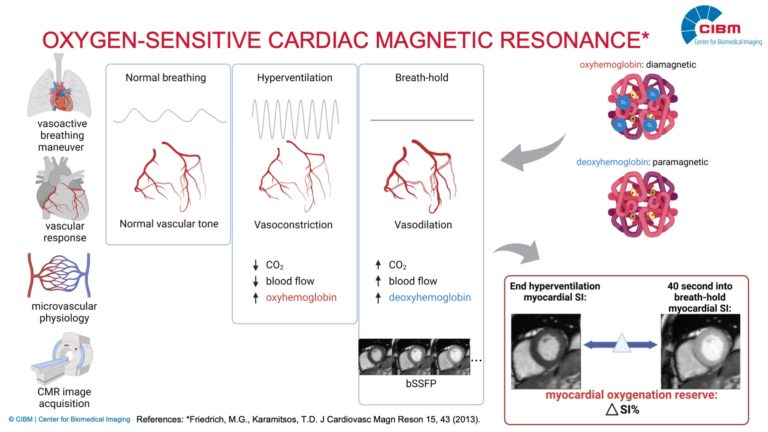
Description: Magnetic resonance imaging enables a safe, non-invasive, assessment of the heart without radiation. It can quantify blood flow, function, and tissue characteristics. However, significant aspects of cardiac MRI have remained unchanged over the past decades, highly specialized personnel is needed, and acquisition is time consuming. To remove these hurdles, we propose to break away from the decades-old paradigm where data are collected in a prospectively triggered fashion, and propose instead to sample image data continuously and irrespective of the heart’s position and contractile state. This can be achieved by combining in depth knowledge of Mathematics, Engineering and Physics. This research will lead to shorter exams for the patient and to more detailed information about the heart muscle and its vessels for the clinician. This translational and interdisciplinary research is conducted in strong collaboration with medical professionals at the CHUV. 

Investigators : Matthias Stuber (CHUV), Jürg Schwitter (CHUV)

Collaborators: Karolinska Institute (Sweden), Emory University (United States of America), Northwestern University (United States of America), Mie University (Japan), Liryc – L’institut De Rythmologie Et Modélisation Cardiaque (France), Children’s Hospital of Philadelphia (United States of America), Medical University of South Carolina (United States of America).

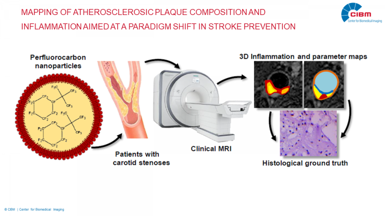
Mapping of atherosclerosic plaque composition and inflammation aimed at a paradigm shift in stroke prevention

Description: Atherosclerosis results in the formation of plaque in the carotid arteries, which can lead to lethal stroke when it ruptures. While surgery and therapy can be used to prevent such rupture, current medical imaging is not specific enough to predict which plaques will rupture. MRI can be used to quantify several rupture risk factors, but not the inflammation severity of the plaque, which is thought to be the most critical risk factor. The goal of this project is therefore to develop a new combined fluorine-19 (19F) MRI and parameter (T1 and T2) mapping technique for the quantification of plaque rupture risk factors: 19F MRI will be used for inflammation mapping, while the parameter mapping will be used for tissue characterization.

Investigator: Ruud van Heeswijk (CHUV)

Collaborators: Matthias Stuber (Département de radiologie médicale CHUV), Samuel Rotman (Institut Universitaire de Pathologie CHUV), Francois Saucy  (Service de Chirurgie Cardio-Vasculaire Département de chirurgie, CHUV), Patrik Michel (Service de Neurologie Département des Neurosciences Cliniques CHUV), Thierry Buclin,  (Service de Pharmacologie clinique Département Médecine Laboratoire Pathologie, CHUV)

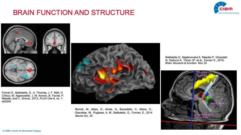
Neuro MRI

Description: Coming soon…

Investigator: Eleonora Fornari (CHUV)

Collaborators: Coming soon…