CIBM MRI EPFL SECTION
MR Imaging Technology
Section Head: Prof. Dimitrios Karampinos (EPFL)
The activities of the CIBM MRI EPFL MR Imaging Technology Section focus on the development and application of state-of-the-art MR imaging methodologies to advance modern clinical diagnostics and to address questions in understanding healthy human physiology, human development and disease pathophysiology. As the applications of MRI continue to expand, our multidisciplinary team is committed to developing and sharing technological solutions that make MRI more robust, objective, efficient and informative in its application.
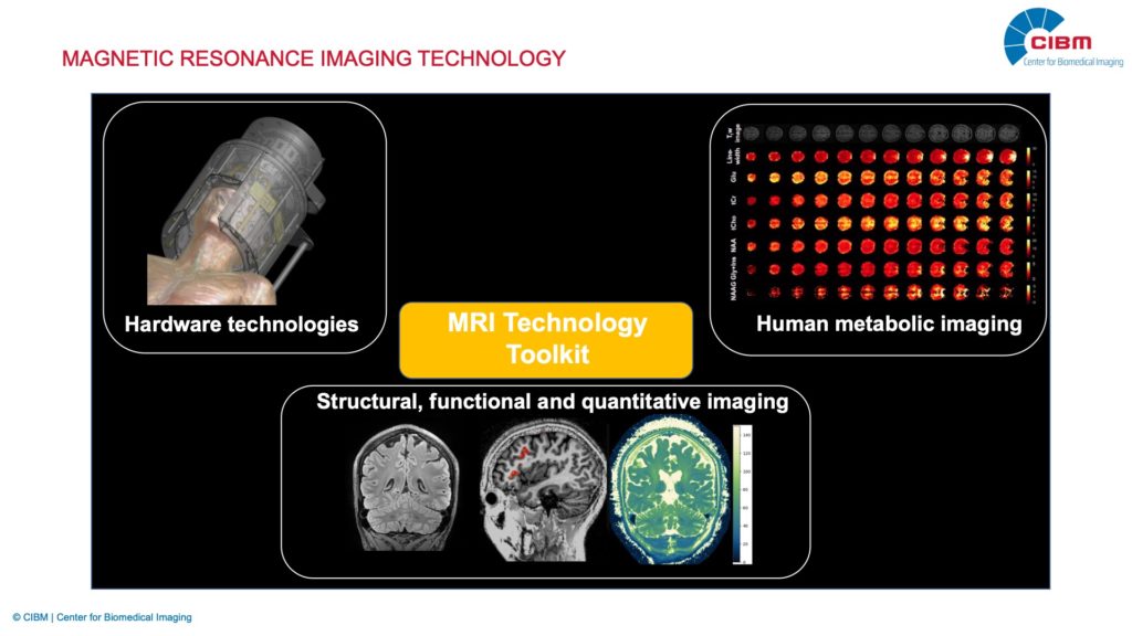
Our methodological toolkit encloses three main components:
- Novel hardware technologies.
- Advanced methodology and (multimodal) applications for structural, functional and quantitative imaging.
- Tools for state-of-the-art metabolic imaging.
A strong focus of the current research activities is on neuroimaging especially at high magnetic field strength, but our activities are continuously growing across field strengths and anatomies.
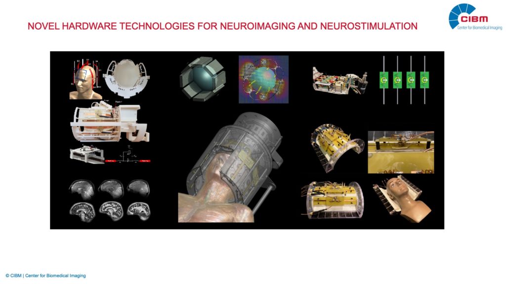
Hardware technologies
High receive sensitivity is the holy grail of every magnetic resonance imaging (MRI) study. Our research aims to substantially boost receive sensitivity in different neuroimaging applications, accelerating total scan time and increasing spatial resolution. We develop innovative hardware concepts for the next generation of MRI detectors, particularly suitable for ultra-high field (UHF) MRI (B0 ≥ 7 T), focusing on unconventional transmit and receive radio frequency (RF) coil arrays involving strategies such as dipole antennas, dielectric resonators, and flexible loop coils. In addition, we explore new solutions for advanced, MRI-based techniques that can provide unique insights into the metabolism and function of the brain, such as phosphorus (31P) MR spectroscopy and electroencephalography combined with functional MRI (EEG/fMRI). Furthermore, we are working on highly promising, non-invasive human brain stimulation strategies. In particular, we develop hybrid hardware systems enabling transcranial low-intensity focused ultrasound (LIFU) neuromodulation combined with UHF-MRI, aiming at finding new ways to treat neurological and neuropsychiatric diseases.
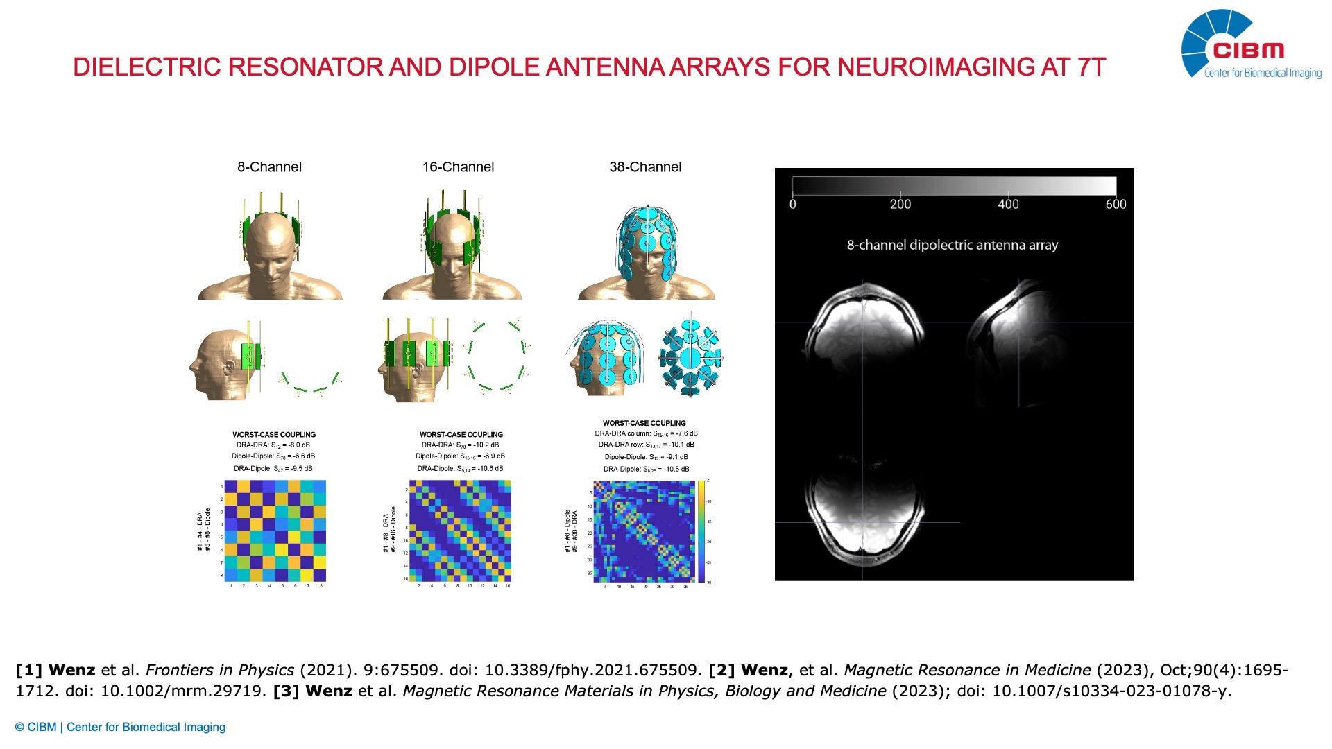
Dielectric resonator and dipole antenna arrays for neuroimaging at 7T
Description: Dielectric resonator antenna (DRA) arrays are a promising alternative for MRI at 7T, which require no additional decoupling circuits and only a minimal number of lumped components. DRAs can be manufactured using ceramic materials with an extremely high dielectric constant (up to thousands). We combine dielectric resonator antennas with dipole antennas to increase SNR in the deep brain in neuroimaging at 7T, which is challenging. Furthermore, we want to benefit from a remarkably strong magnetic field near DRAs and substantially boost SNR in peripheral brain areas.
Investigator: Daniel Wenz (EPFL)
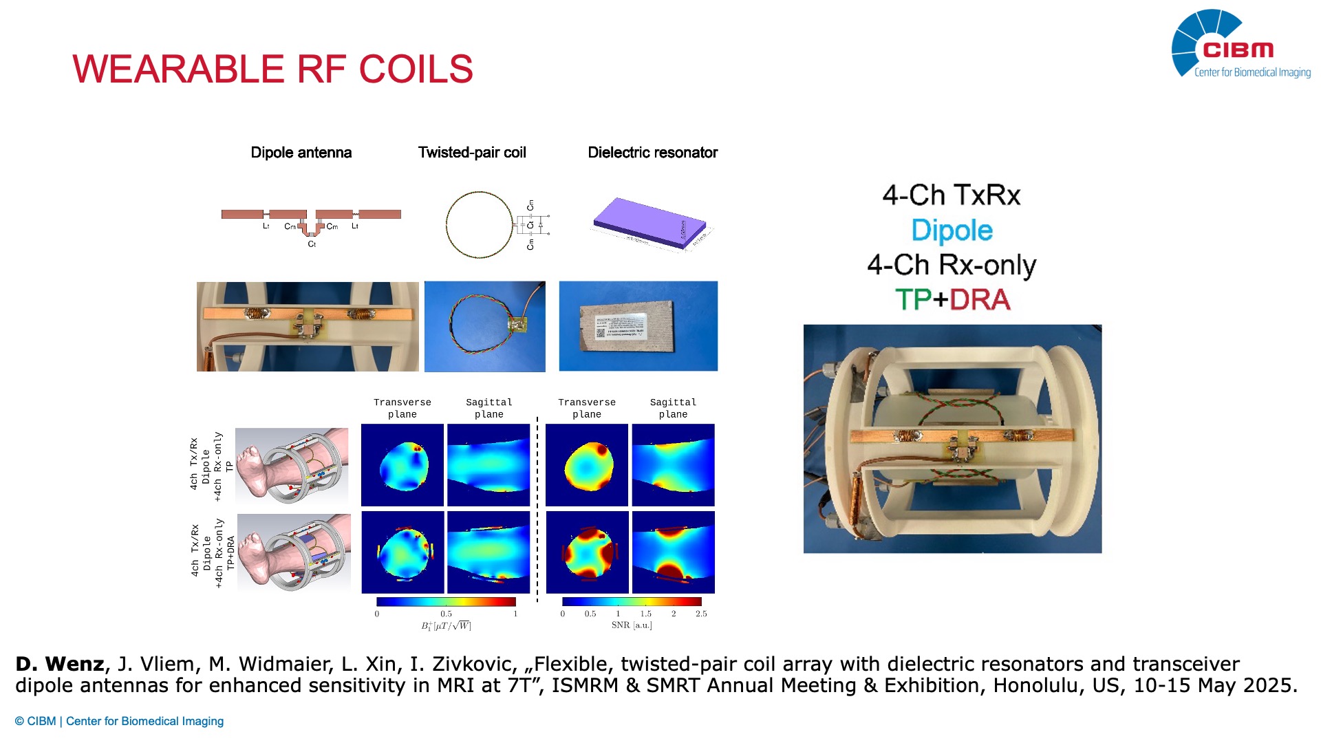
Wearable RF coils
Description: Bringing RF coils closer to the head can substantially increase SNR in brain MRI. To achieve this goal, we need flexible, decoupled multi-channel receive arrays. We study twisted-pair coil arrays and aim to employ them as wearable MRI detectors for applications such as pediatric MRI and simultaneous EEG/fMRI. We also investigate how the performance of twisted-pair coils can be further boosted by combining them with other complementary strategies, such as high-permittivity materials (HPMs) and dipole antennas.
Investigator: Daniel Wenz (EPFL)
Collaborators: Irena Zivkovic (Technical University of Eindhoven, The Netherlands), Frederic Grouiller (UNIGE)
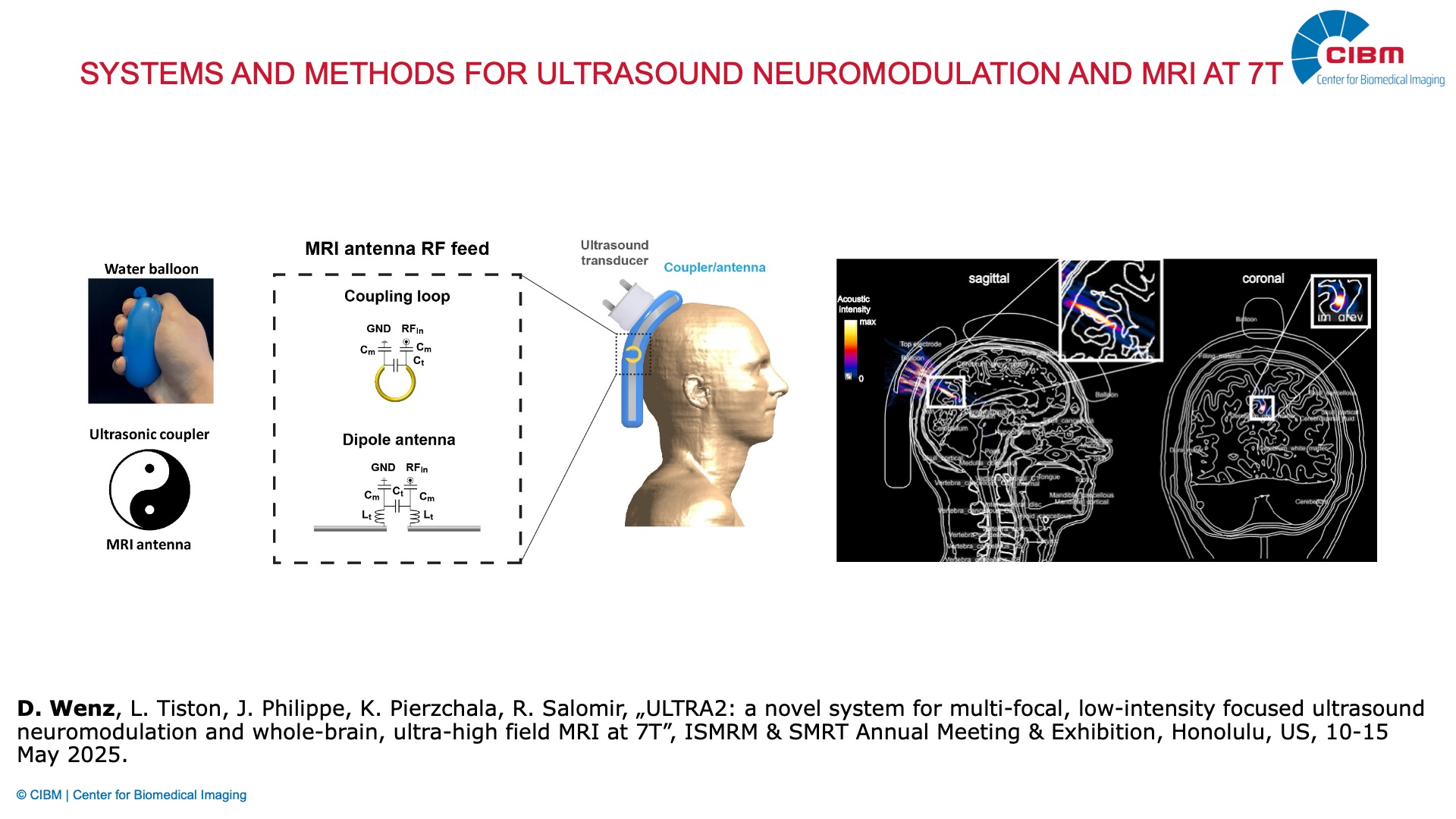
Systems and methods for ultrasound neuromodulation and MRI at 7T
Description: Transcranial low-intensity focused ultrasound (LIFU) is an emerging technique for non-invasive brain stimulation. LIFU can provide remarkably high precision, selectivity, and penetration depth. We develop advanced solutions to ensure excellent performance in simultaneous, multi-focal LIFU neuromodulation and whole-brain MRI at 7T. In particular, we exploit the intrinsic duality of acoustic couplers, which can provide efficient LIFU propagation and act as a multi-channel dielectric resonator antenna array.
Investigator: Daniel Wenz (EPFL)
Collaborators: Rares Salomir (HUG, UNIGE), Henrik Odéen (University of Utah, USA)
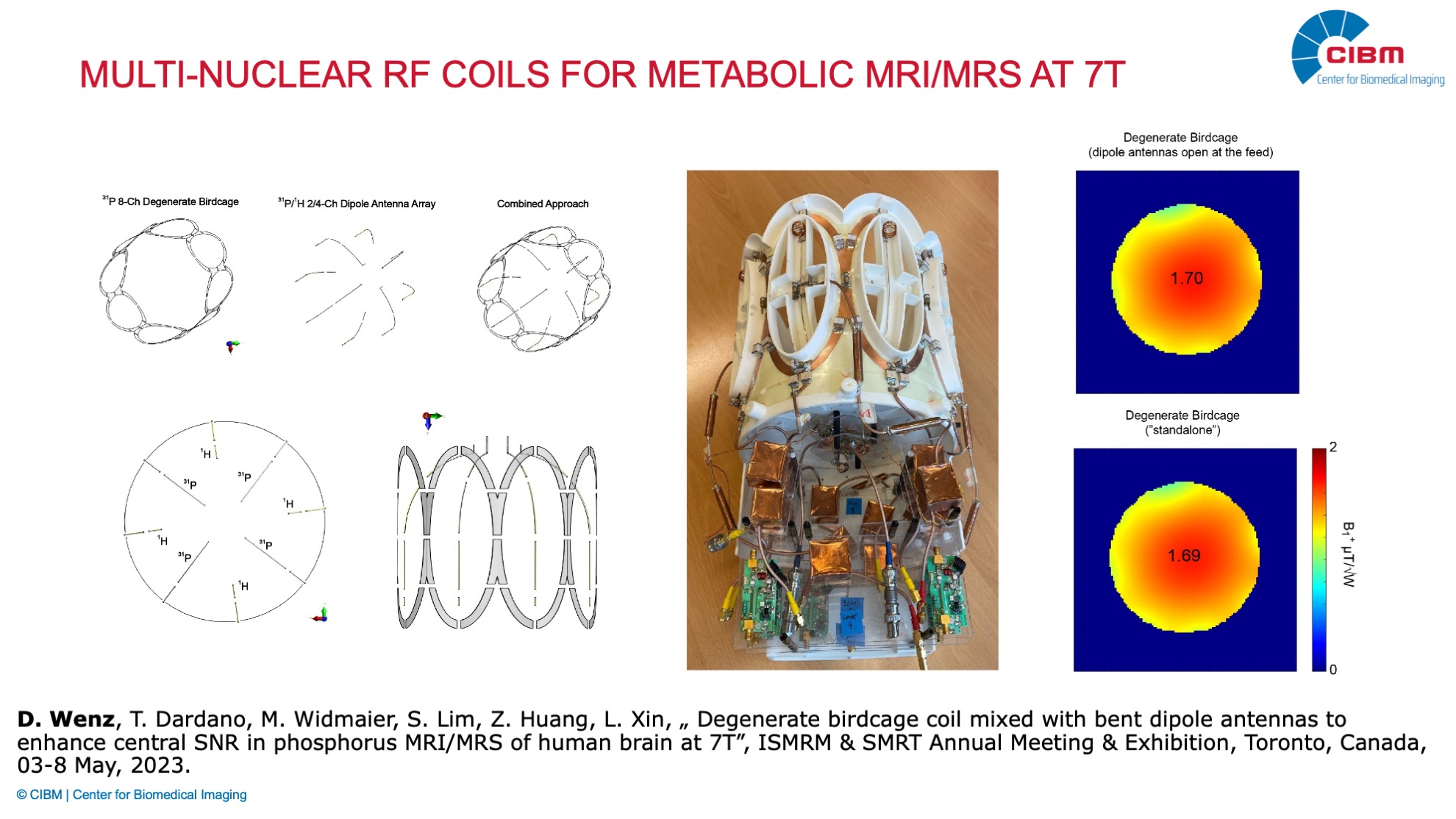
Multi-nuclear RF coils for metabolic MRI/MRS at 7T
Description: Non-proton MRI/MRS involving nuclei, such as phosphorus (31P), is a powerful tool for probing brain metabolism and physiology in vivo. However, SNR in deep brain areas, which are particularly relevant for research in neuropsychiatry, is still unsatisfactory. To address this, we develop new arrays and combine loop elements with dipole antennas, which can better approach the RF current distribution predicted by the ultimate intrinsic SNR theory. In addition, we explore novel, free-of-lossy decoupling circuits strategies that would provide excellent performance (at least 32 channels per nucleus) of both receive arrays for anatomic (1H) and metabolic (31) MRI and MRS.
Investigator: Daniel Wenz (EPFL)
Collaborator: Lijing Xin (EPFL)
Structural, functional and quantitative imaging
Our research is dedicated to the advancement of structural, functional, and quantitative MRI techniques with a strong present focus on 7T MRI. Central to our mission is a strong and ongoing collaboration with industry partners, enabling the co-development of pulse sequence innovations. At 7T MRI, we focus on the design and implementation of optimized imaging protocols and advanced acquisition strategies to address the inherent challenges of 7T MRI—such as field inhomogeneities and SAR constraints. At 3T MRI, we are developing novel methods for quantitative body imaging. By maintaining close and continuous interactions with clinical teams, we ensure that our technological developments are directly translatable to patient care, facilitating the integration of cutting-edge imaging solutions into clinical and research workflows.
RESEARCH TOPICS
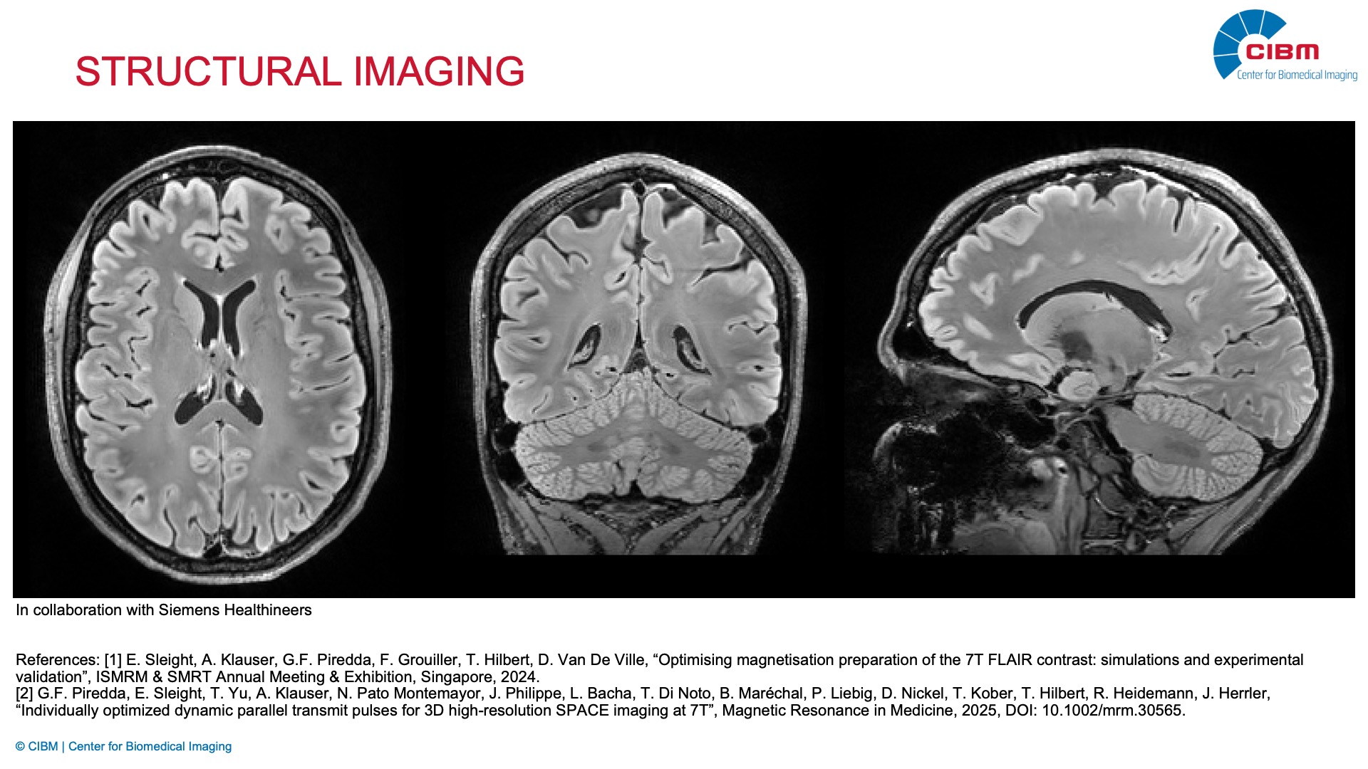
Structural imaging
Description: We have developed high-resolution structural imaging protocols tailored for clinical applications at 7T, with a particular focus on optimising contrast and mitigating field-related artifacts. In collaboration with Siemens Healthineers, we refined the FLAIR sequence to improve lesion detection and ensure robust performance in the presence of B1 inhomogeneities—key challenges in ultra-high field MRI. These advancements have enabled more reliable clinical assessments, particularly in neurological applications.
Investigator: Emilie Sleight (EPFL)
Collaborators: Gian Franco Piredda, Antoine Klauser, Jürgen Herrler, Tom Hilbert (Siemens Healthineers), Felix Kurz (HUG), Frédéric Grouiller (HUG,UNIGE)
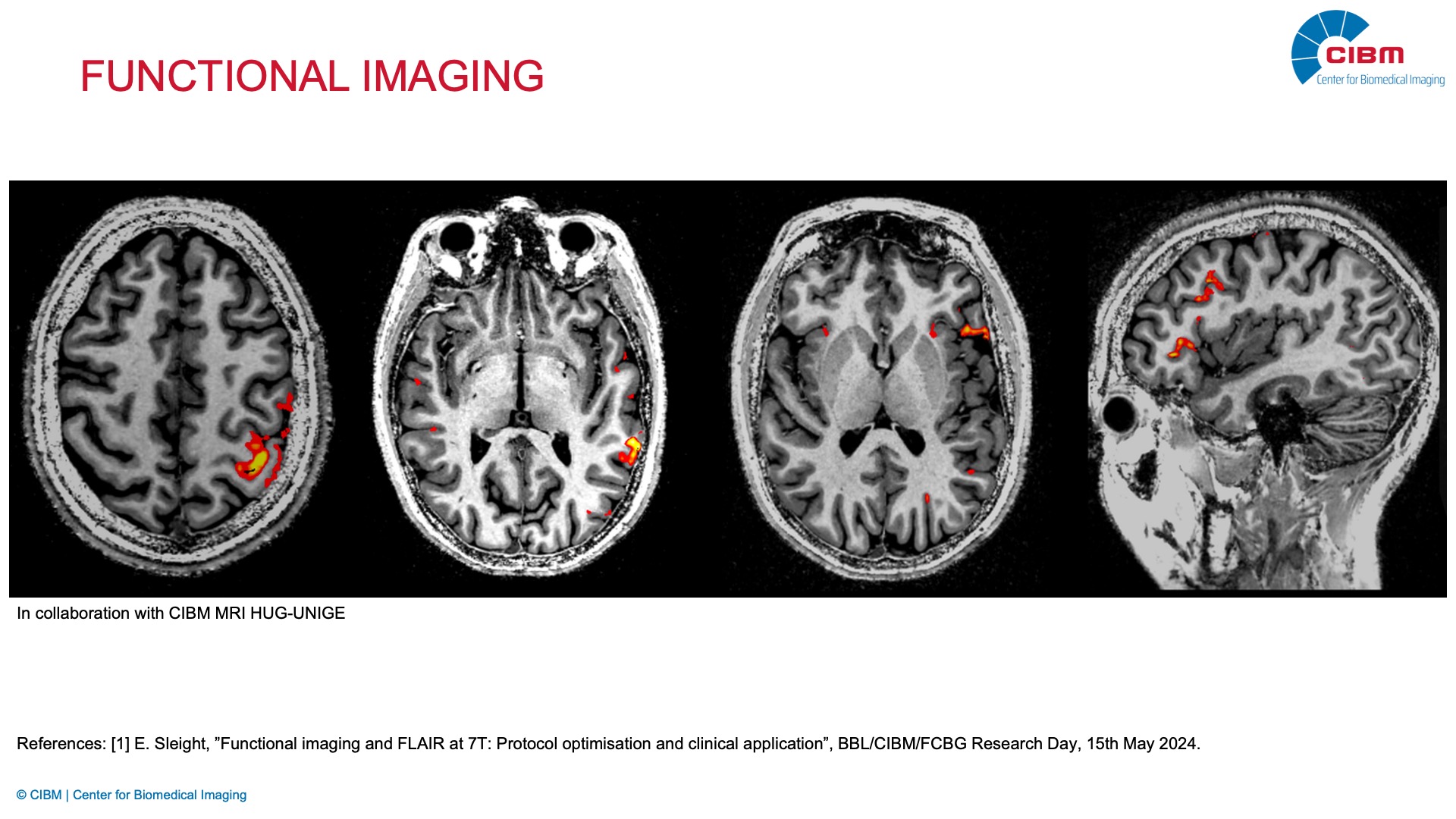
Functional neuroimaging
Description: Our work in functional imaging at 7T focuses on both clinical and research applications. In partnership with the CIBM MRI HUG-UNIGE, we developed specialised 7T fMRI protocols for pre-surgical brain mapping, providing enhanced spatial resolution and sensitivity compared to lower field strengths. In parallel, we are actively engaged in advancing laminar fMRI, aiming to resolve activation patterns across cortical layers and contribute to a deeper understanding of neurovascular coupling and cortical processing at ultra-high resolution.
Investigators: Emilie Sleight (EPFL), Frédéric Grouiller (HUG,UNIGE)
Collaborators: Sébastien Courvoisier, Giannina Rita Iannotti (HUG,UNIGE), Karolis Degutis (EPFL)
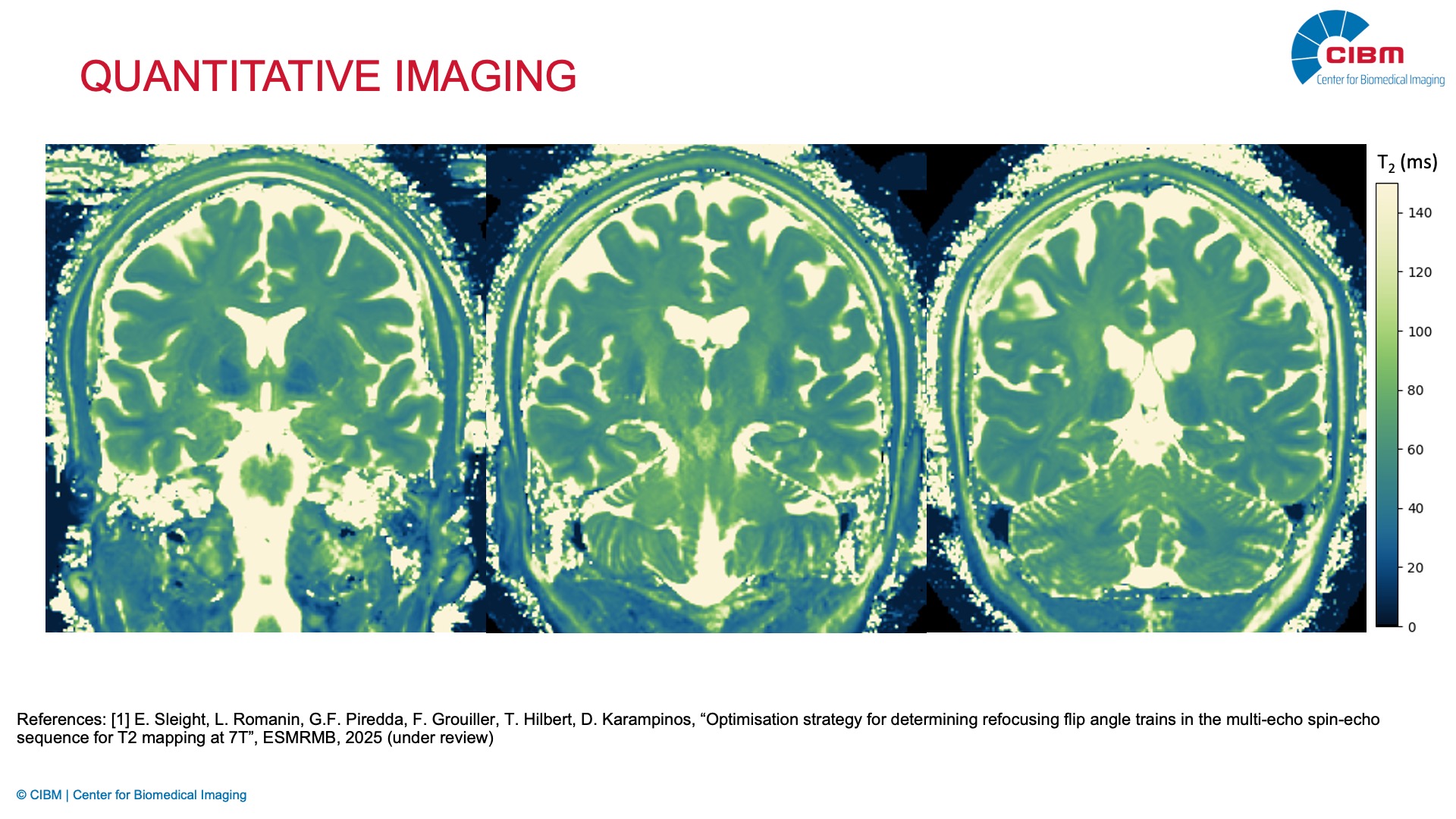
Quantitative imaging
Description: We are actively developing quantitative imaging techniques at 7T, including T2 mapping and other parametric mapping methods, to provide objective and reproducible biomarkers of tissue properties. Our work addresses key technical challenges such as increased specific absorption rate and field inhomogeneity, leveraging novel pulse sequence designs. These efforts are essential for enabling reliable and standardised quantitative imaging in both clinical and research settings.
Investigators: Dimitrios Karampinos, Emilie Sleight (EPFL)
Collaborators: Gian Franco Piredda, Ludovica Romanin, Tom Hilbert (Siemens Healthineers)
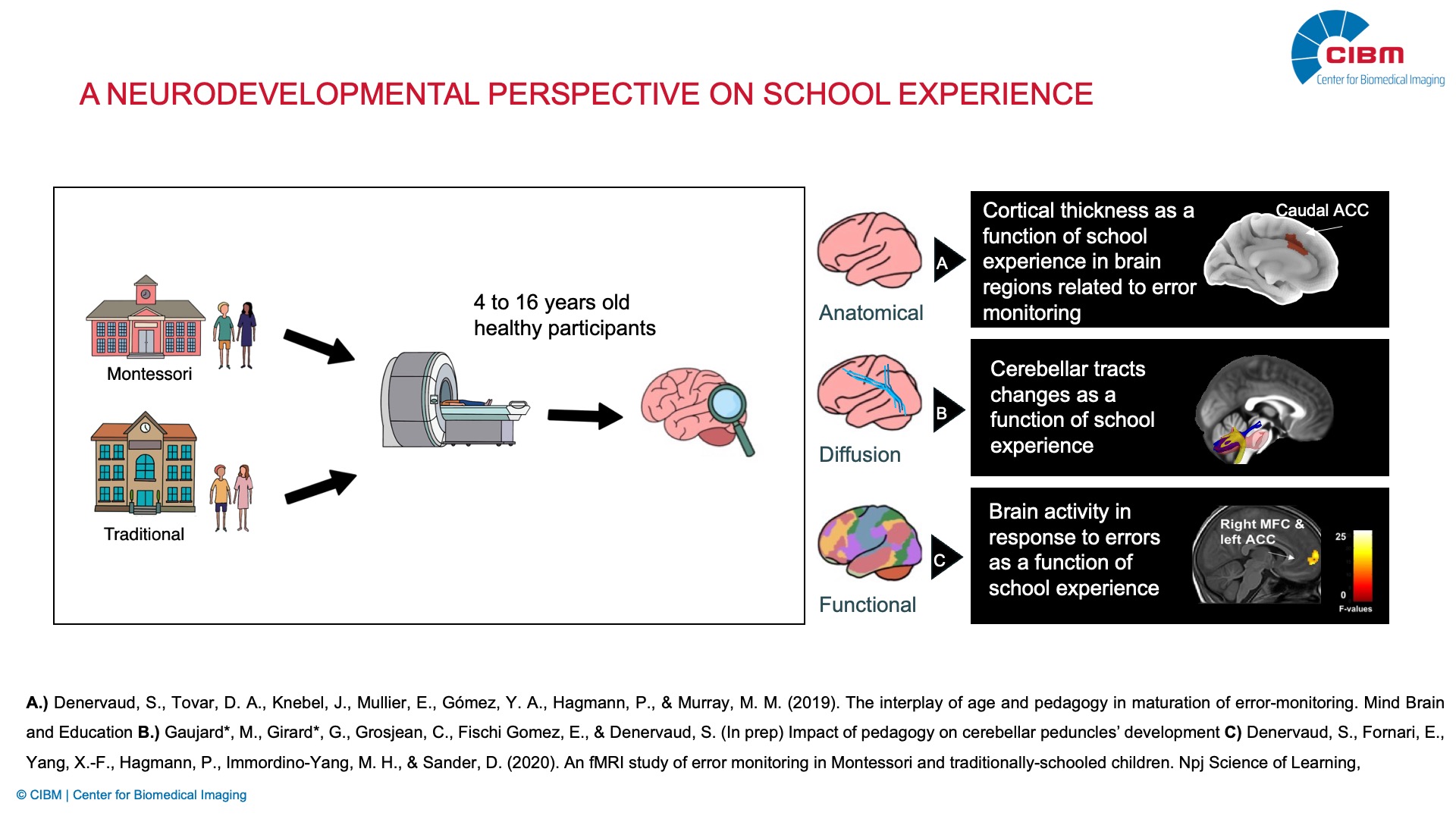
Neurodevelopmental imaging
Description: This research explores how different educational environments shape the developing brain, focusing on the neural mechanisms underlying creativity, adaptability, and resilience in children and adolescents. By combining advanced MR imaging techniques with complementary neuroimaging approaches such as EEG and behavioral assessments, the project investigates how early life experience such as schooling influence healthy cognitive and emotional development.
The study applies structural, functional, and quantitative MRI to examine brain regions involved in error monitoring, cognitive flexibility, reward processing, and social interaction. By integrating these insights with real-world educational practices, the research aims to develop a neurobiological framework for optimizing learning environments, with far-reaching implications for both educational policy and child well-being.
Investigator: Solange Denervaud (EPFL)
Collaborators: Merixtell Bach Cuadra (CHUV-UNIL), Eleonora Fornari (CHUV), Gabriel Girard (Université de Sherbrooke), Gustavo Deco (Universitat Pompeu Fabra), Mary Helen Immordino Yang (USC), Beata Bednarczuk (Maria Curie-Skłodowska University)
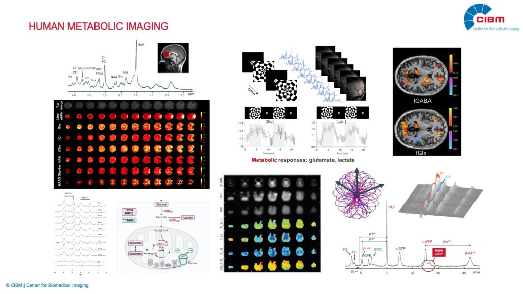
Human metabolic imaging
Our research focuses on innovation in multinuclear magnetic resonance methodologies for metabolic imaging in humans. We develop accelerated acquisition strategies and advanced reconstruction methods to reduce scan times for both static and dynamic mapping of metabolite levels at the clinical MRI platform. Additionally, we are establishing and expanding unique X-nuclei imaging capabilities for clinical applications, including 31P, 13C, and extending to 2H and other nuclei. Our human metabolic imaging research offers the following advanced methodologies:
- Neurochemical profiling and imaging with 1H magnetic resonance spectroscopy and spectroscopic imaging (e.g. neurotransmitters: Glutamate and GABA; and energy metabolite Lactate)
- Novel functional MR imaging method for mapping brain functional-metabolic connectivity
- Whole-brain mapping of ATP metabolism (ATP synthase and creatine kinase) by 31P magnetic resonance fingerprinting (MRF)
- Mapping intracellular pH, NAD, phosphomonoesters, phosphodiesters, and phosphocreatine by 31P MR spectroscopic imaging
- Neuronal and astrocytic oxidative metabolism by dynamic 13C magnetic resonance spectroscopy
We leveraging these cutting-edge techniques to gain insights into brain function and neuropsychiatric disorders, elucidating underlying molecular mechanisms, and ultimately uncovering novel biomarkers for early and precise diagnosis and treatment.
RESEARCH TOPICS
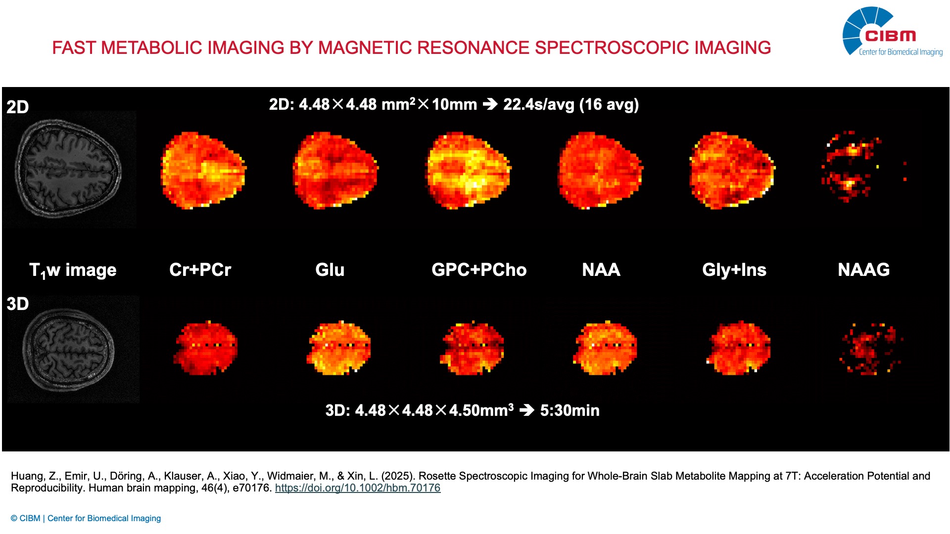
Fast metabolic imaging by magnetic resonance spectroscopic imaging
Description: Whole-brain proton magnetic resonance spectroscopic imaging (1H-MRSI) is a non-invasive technique for assessing neurochemical distribution in the brain, offering valuable insights into brain functions and neural diseases. It greatly benefits from the improved SNR at ultrahigh field strengths. However, 1H-MRSI still faces challenges, such as long acquisition time and signal contamination from water and lipids. In this project, we develop 2D and 3D short TR/TE 1H-FID-MRSI sequences using rosette trajectories with high spatial resolution for mapping brain metabolites including N-Acetyle-L-aspartic acid (NAA), Glutamate (Glu), total choline, Creatine and Phosphocreatine (tCr), and Glycine and myo-Inositol (Gly+Ins).
Investigators: Lijing Xin (EPFL), Andre Doring (EPFL), Zhiwei Huang (EPFL)
Collaborators: Uzay Emir (UNC, USA), Zhi-Pei Liang (UIUC, USA), Antoine Klauser (Siemens)
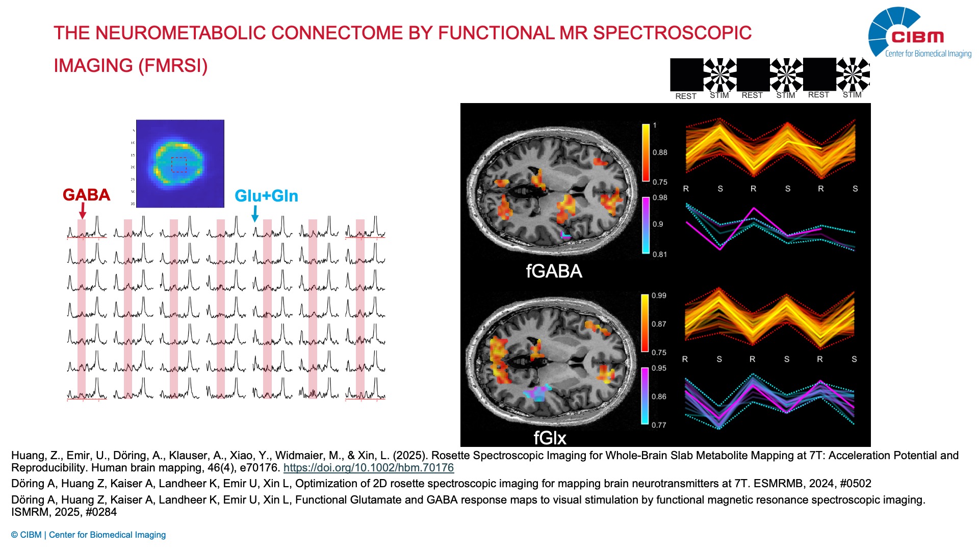
The neurometabolic connectome by functional MR spectroscopic imaging (fMRSI)
Description: Magnetic resonance spectroscopy (MRS) is a unique non-invasive technique to measure a variety of brain metabolites (e.g., N-acetylaspartate, creatines, choline, inositol) and certain neurotransmitters (glutamate, Gamma-Aminobutyric Acid). Functional MRS (fMRS) allows us to monitor the metabolites dynamics from rest to neuronal activation. However, so far it has remained mostly a single voxel technique with limited spatial and temporal resolution. By leveraging novel MR sequence designs (edited-MRSI, FID-MRSI), spatial encoding schemes (rosette trajectory), and reconstruction methods (compressed sensing) on a state-of-the-art 7T Terra.X MR scanner, the aim of this project is to improve spatial and temporal resolution to establish functional MR spectroscopic imaging (fMRSI) that enables the dynamic measurement of modulation patterns of neurotransmitter concentrations upon stimulation (sensory stimulations and cognitive tasks) in the human brain, and then mapping of brain metabolic connectivity.
Investigators: Lijing Xin (EPFL), Andre Doring (EPFL), Zhiwei Huang (EPFL), Antonia Kaiser (EPFL)
Collaborators: Uzay Emir (UNC, USA), Dimitrios Karampinos (EPFL), Zhi-Pei Liang (UIUC, USA), Martin Zach, Michael Unser, Dimitri Van de Ville (EPFL)
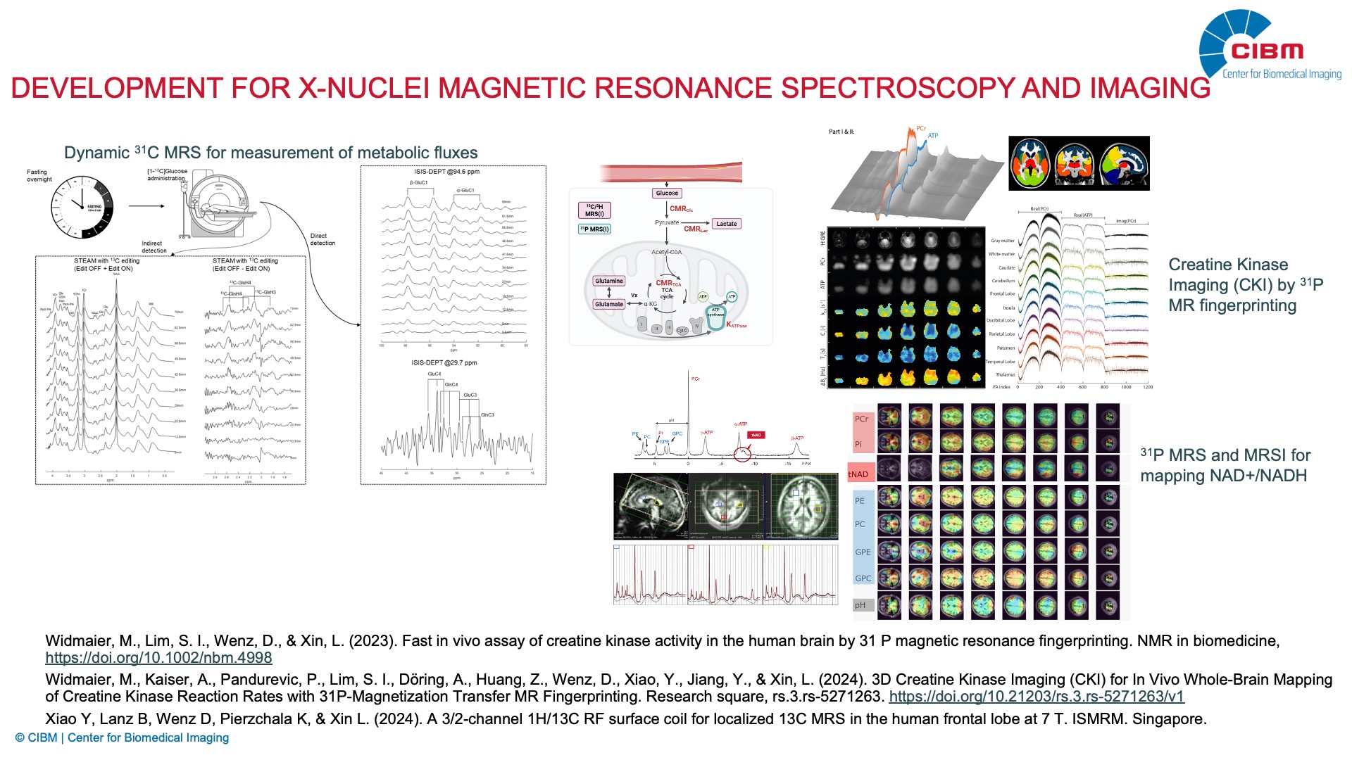
Method development for X-nuclei magnetic resonance spectroscopy and imaging
Description: X-nuclei magnetic resonance spectroscopy (MRS) allows non-invasive measurement of neurochemical, metabolic and physiological events in the living brain. The increase in sensitivity and resolution is currently driving the use of ultra-high magnetic field scanners. However, to fully exploit these advantages of an ultra-high field human scanner necessitates the development first of advanced methodologies. Especially, the X- nuclei MRS functionality of clinical scanners is still far behind magnetic resonance imaging techniques and that on preclinical scanners, due to the need to overcome multiple technical challenges. Therefore, we focus on surmounting the technical issues, developing new X-nuclei MRS methods and advancing their capability for human MR scanner at high magnetic field. Currently our focuses are the metabolic fluxes (TCA cycle metabolism and ATP metabolism) measurement using dynamic 13C MRS, 31P MRS, MRSI and MR fingerprinting.
Investigators: Lijing Xin (EPFL), Mark Widmaier (EPFL), Ying Xiao (EPFL), Daniel Wenz (EPFL)
Collaborators: Uzay Emir (UNC, USA), Yun Jiang (University of Michigan, USA), Xin Yu (Case Western Reserve University, Cleveland, USA), Bernard Lanz (EPFL).
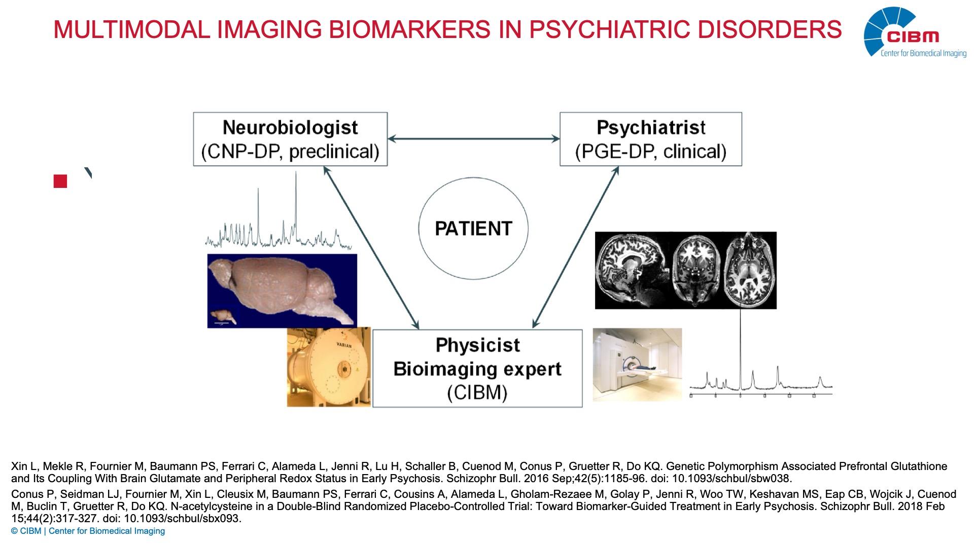
Multimodal Imaging Biomarkers in Psychiatric Disorders
Description: Psychiatric disorders are complex neurodevelopmental disorders involving interplay between genetic, developmental and environmental factors. Using advanced 1H, 13C and 31P MRS techniques, we carry on a translational approach in patients with psychosis, for the exploration of potential biomarkers and the identification of molecular mechanisms affecting brain development and regulation that are relevant for the pathophysiology of schizophrenia. The discovery of biomarkers will help us to achieve early diagnose of the disease and ultimately early intervention or even prevention.
Investigators: Lijing Xin (EPFL), Antonia Kaiser (EPFL)
Collaborators: Kim Q Do (UNIL), Philippe Conus (Service of General Psychiatry, Department of Psychiatry, CHUV), Ines Khadimallah (Center for Psychiatric Neuroscience, Department of Psychiatry, CHUV), Patric Hagmann (Department of Radiology, CHUV), Paul Klauser (Center for Psychiatric Neuroscience, Department of Psychiatry, CHUV), Stefan Kaiser (Geneva University Hospital), Matthias Kirschner (Geneva University Hospital), Indrit Bègue (Geneva University Hospital).
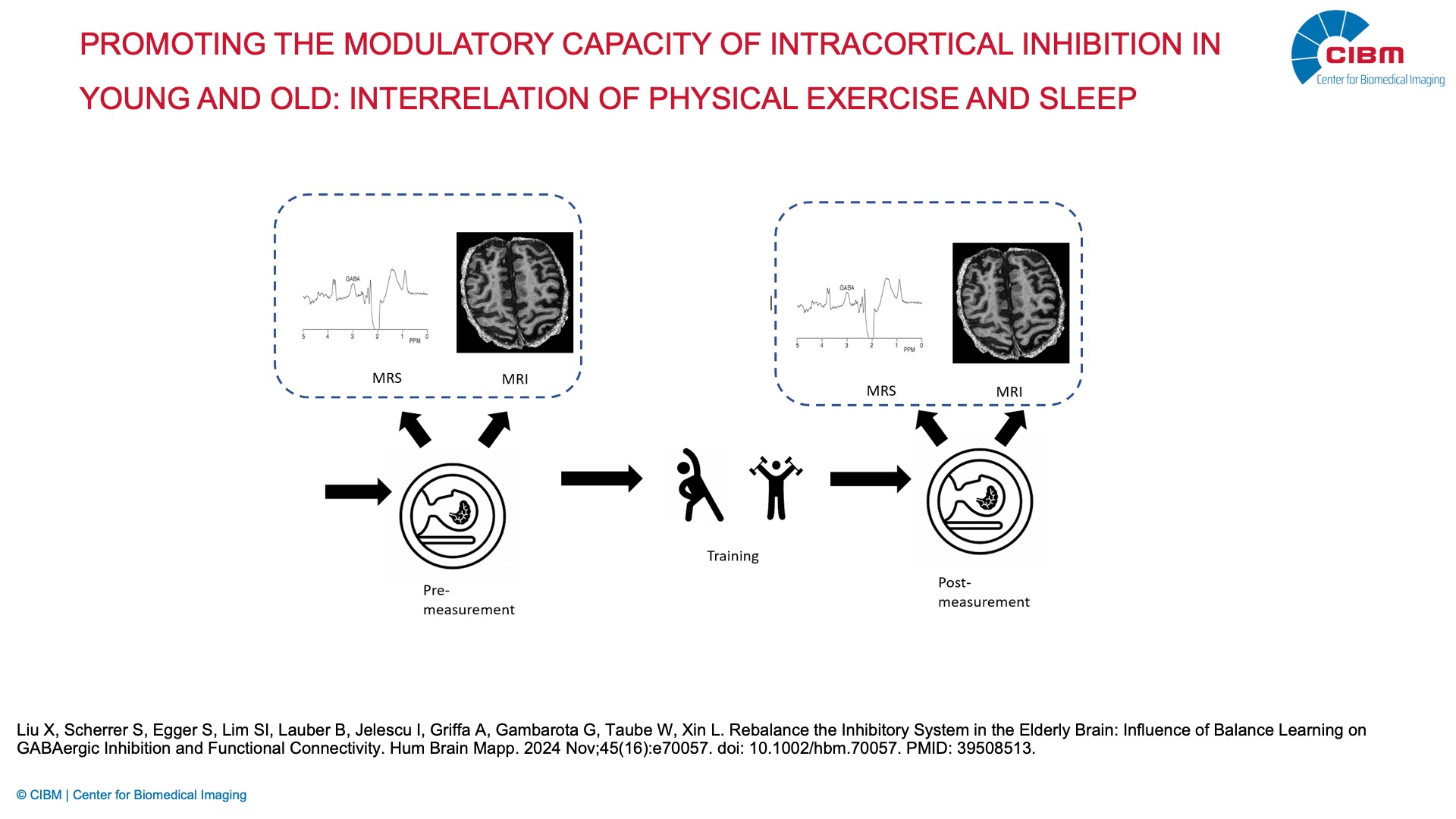
Promoting the modulatory capacity of intracortical inhibition in young and old: interrelation of physical exercise and sleep
Description: The human cortical inhibitory system is known to play a crucial role for normal brain development, function and plasticity thereby acting on both, cognitive and motor processes. Furthermore, cortical inhibition is crucial for sleep induction and sleep maintenance. The principal inhibitory neurotransmitter in the central nervous system is gamma-aminobutyric acid (GABA). The general amount of GABAergic inhibition is crucial with respect to motor control and motor learning but rather the capacity to task- and phase-specifically modulate GABA release, i.e. the modulatory range. Furthermore, modulation of GABA release is also vital for sleep induction and sleep maintenance. The aim of the current study is to investigate the close reciprocal interrelation of physical activity and sleep on the mechanistic and behavioral level. This could be of great significance as there is growing recognition of the importance of sleep to improve population health due to the convincing evidence linking sleep to a range of health outcomes.
Investigators: Lijing Xin (EPFL), Wolfgang Taube (UNIFR)
Collaborator: Benedikt Lauber (UNIFR)