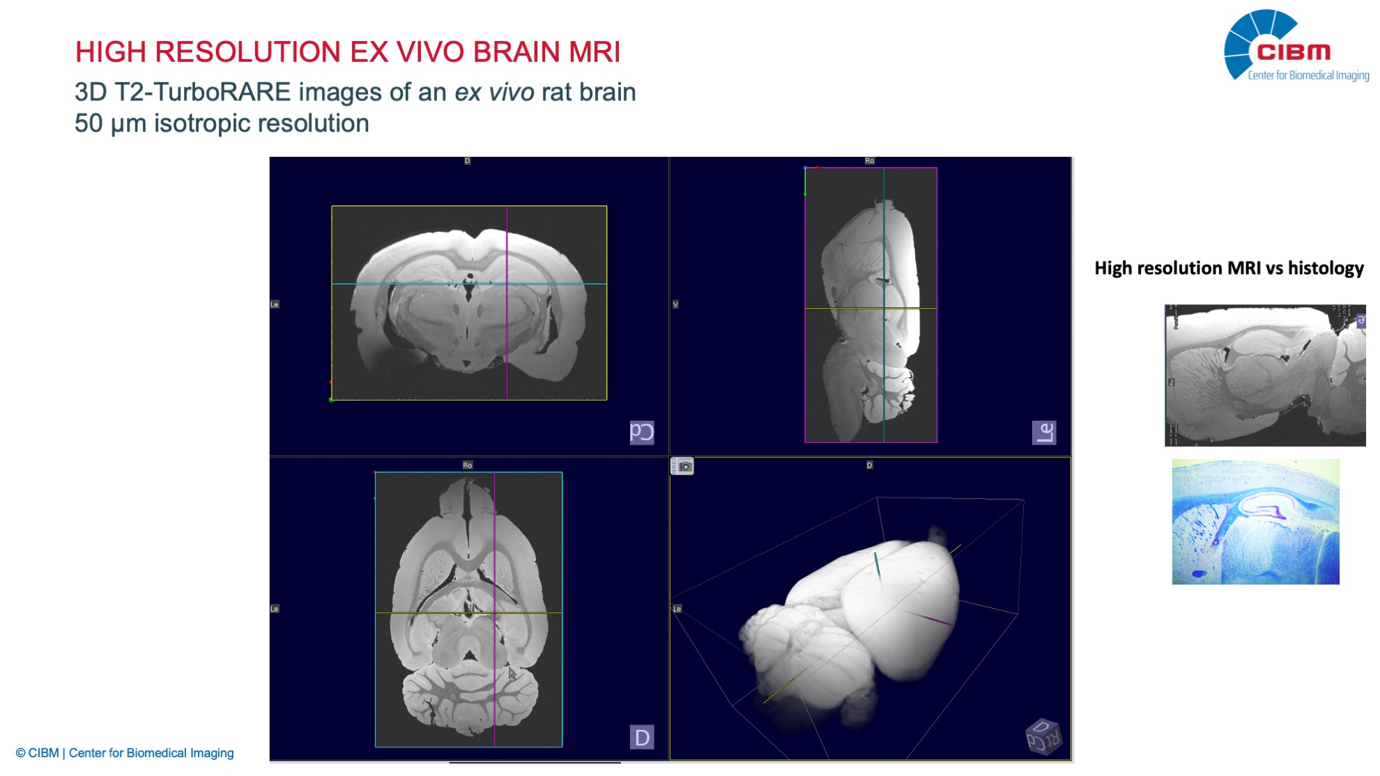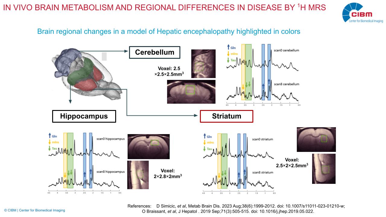
Mapping brain microstructure in vivo using diffusion MR Spectroscopy and MR Imaging
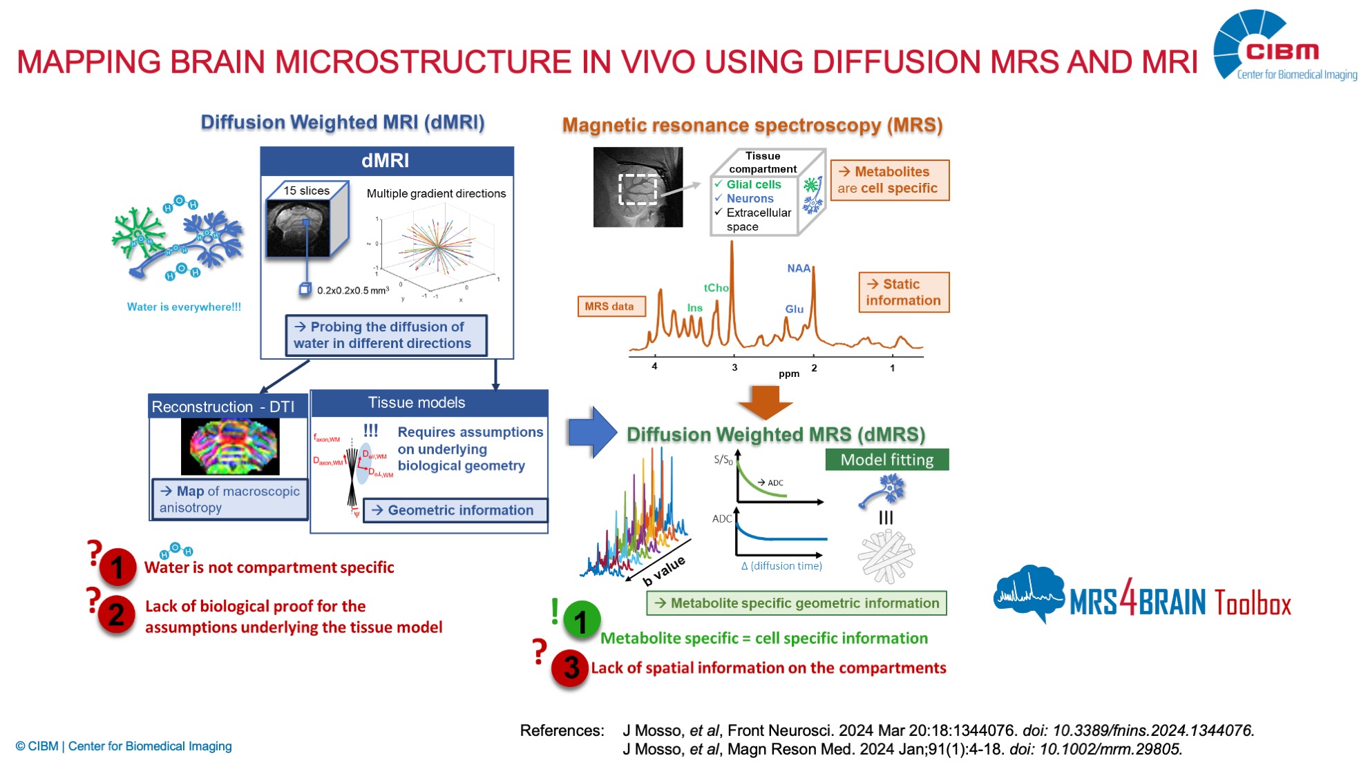
Description: The temporal and regional evolution of the brain during development, disease or injury is characterized by numerous microstructural and metabolic changes, for which new non-invasive methods advancing the quantification of brain microstructure are necessary. Diffusion MRI (dMRI) of water provides information about tissue microstructure non-invasively, but its specificity is limited due to ubiquitous presence of water. The biggest challenge remains the validation of the biological accuracy of estimated parameters. In contrast, brain metabolites measured by MR spectroscopy (MRS) are predominantly intracellular and some have a reportedly preferential localization in specific brain cell types. Diffusion MRS (dMRS) can thus provide quantification of cell-type specific microstructure. Taking advantage of this specificity to tag microstructure across the brain, however, still requires thorough validation.
Our overall goal is therefore to push further a new concept for quantification and validation of brain microstructure through diffusion of brain metabolites and water at cellular and sub-cellular scale, by implementing an in vivo innovative multi-modal approach combining novel dMRS acquisitions, state-of-the-art postprocessing and metabolite diffusion modeling techniques validated with 2-photon microscopy and mass spectrometry. Our target application is the critical period of brain development and associated potential injury, that we will assess using both 3D brain cell organoids (validation of metabolite diffusion metrics) and in vivo rodent models.
Investigator: Cristina Cudalbu, Tan Toi Phan, Eloise Mougel, Thi Ngoc Anh Dinh, Thanh Phong Lê (EPFL).
Collaborators: Ileana Jelescu (CHUV, UNIL), Julien Valette (CEA, Paris), Olivier Braissant (CHUV, UNIL), Stephane Sizonenko (HUG, UNIGE)