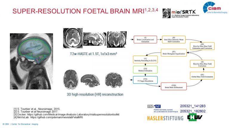
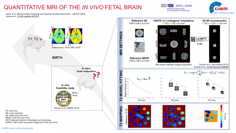
Super-Resolution T2w Foetal Brain MRI for Computer Assisted Diagnosis and Quantitative Analysis
Description: We develop advanced quantitative imaging techniques for studying the maturation of the in-vivo human brain in its early stages of development, when it undergoes the most significant changes. One research line is dedicated to the image super-resolution reconstruction and segmentation of T2w fetal imaging. This will provide accurate quantitative analysis of the growing brain anatomy by morphological biomarkers. Another research line is related to support the translation of the developed reconstruction and segmentation methods to the clinical environment and their evaluation in daily exploration of fetal brain MR images. This research is supported by Swiss National Science Foundation (205321_182602 and 205321_141283), Haslerstiftung and the Radiology Department of the Lausanne University Hospital.
Investigators: Priscille de Dumast (UNIL), Hamza Kebiri (UNIL), Hélène Lajous (UNIL), Meritxell Bach Cuadra (UNIL)
Collaborators: Dr. S. Tourbier (Radiology Department CHUV), Prof. P. Hagmann (Radiology Department CHUV), Prof. P. Maeder (Radiology Department CHUV), Dr V. Dunet (Radiology Department CHUV), Dr. M. Koob (Radiology Department CHUV), Prof. R. Meuli (Radiology Department CHUV), T. Yu (EPFL), Prof. J.-Ph. Thiran (EPFL), Dr. B. Marechal (Siemens Healthcare), M. R. Corredor (Siemens Healthcare)


