CIBM SP CHUV-EPFL SECTION

Computational Medical Imaging

CIBM Section Head: Prof. Jean-Philippe Thiran (EPFL)

The CIBM Signal Processing CHUV–EPFL Computational Medical Imaging Section develops advanced methods for modeling, acquisition, reconstruction, and analysis of (bio)medical images, with a strong emphasis on MRI and ultrasound.

Computational Medical Imaging for Microstructure Characterization

In the MRI part, the research is globally oriented toward microstructure imaging, with applications in neuroscience, oncology, and clinical translation. The ultimate aim is the integration of biologically informed modeling, simulation, and machine learning to bridge the gap between virtual experiments and real-world patient imaging.

RESEARCH TOPICS

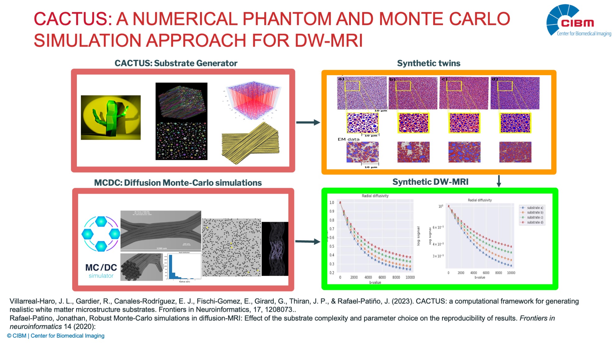
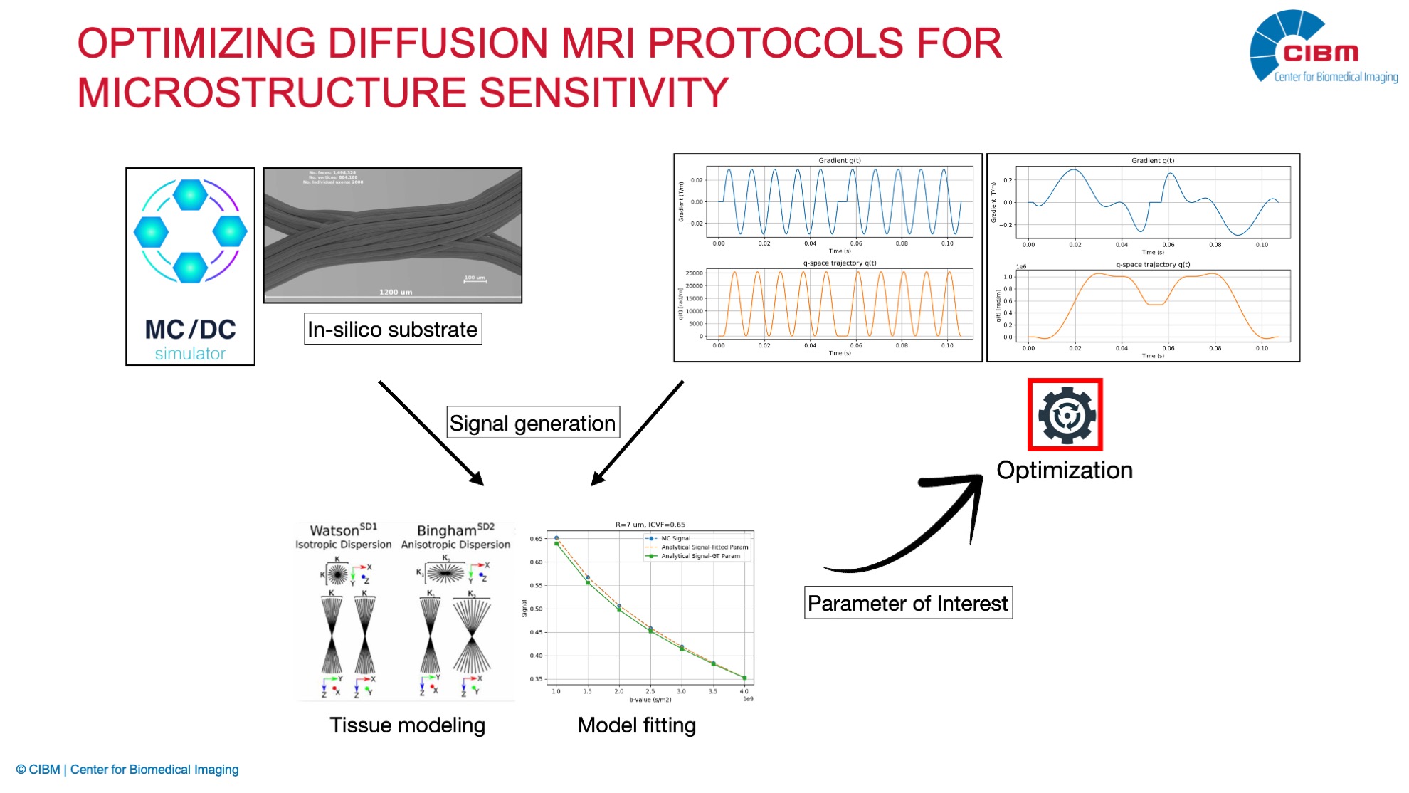
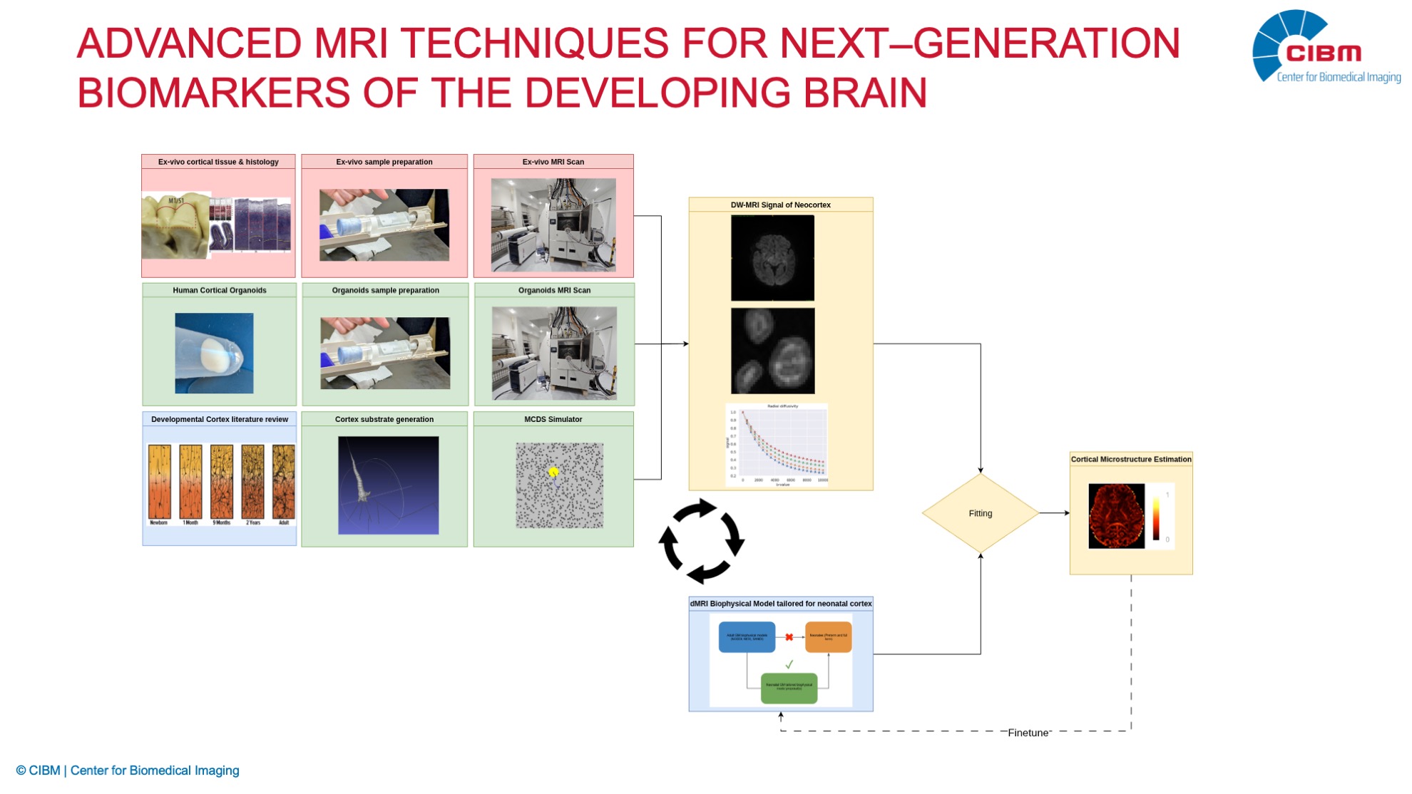
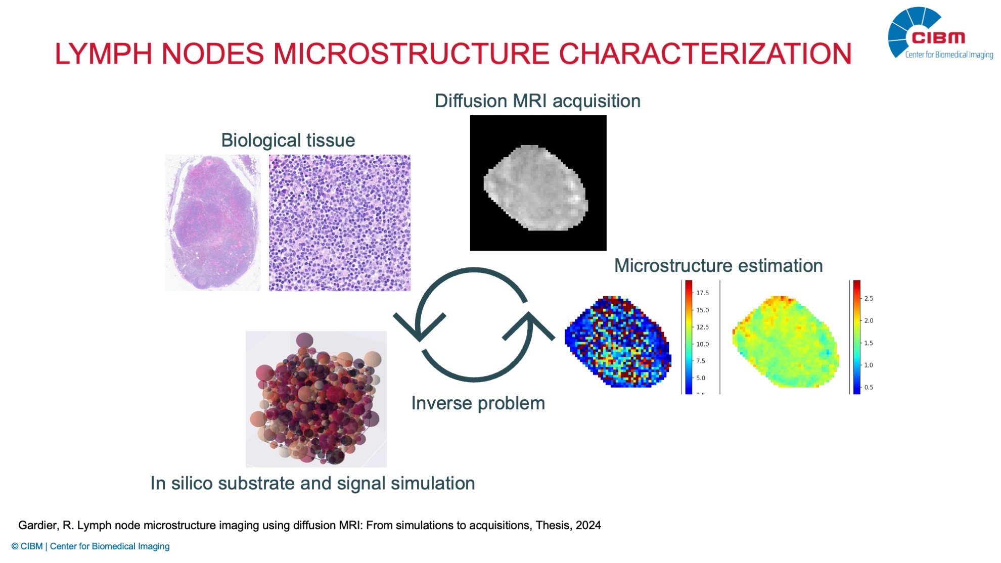
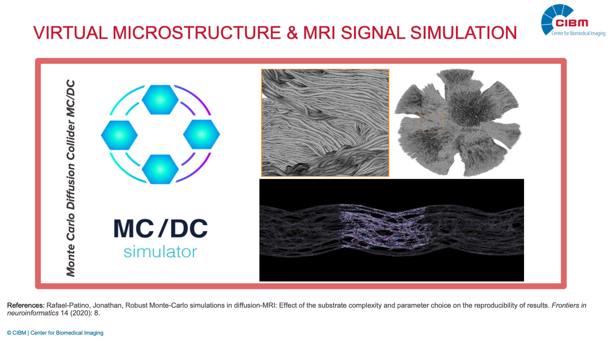
White Matter Microstructure Imaging with CACTUS: A Numerical Phantom and Monte Carlo Simulation Approach for DW-MRI

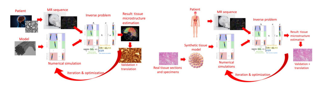
Description: The CACTUS Framework for White Matter Microstructure Imaging aims to create biologically realistic computational substrates of brain and other tissues, combined with high-fidelity Monte Carlo simulations of diffusion MRI. By simulating water diffusion and MR signal formation under advanced acquisition protocols, this project provides a robust platform to design, test, and validate new imaging models with direct applications in both neuroscience and oncology.

Investigator: J.-P. Thiran, J. Rafael-Patiño (EPFL)

CACTUS Numerical Phantoms for DW-MRI

Description: This project aims at leveraging the CACTUS framework to generate highly customizable virtual white matter environments. These phantoms allow researchers to explore how tissue properties such as fiber density, orientation, and tortuosity affect diffusion MRI signals, thereby improving the interpretation of complex brain imaging datasets and advancing our understanding of white matter microstructur.

Investigator: J.-P. Thiran, J.L. Villarreal-Haro (EPFL)

Optimizing Diffusion MRI Protocols for Microstructural Sensitivity

Description: Diffusion MRI has been widely used as noninvasive method to investigate brain microstructure, but conventional protocols often lack specificity, causing different microstructural features to produce indistinguishable signatures. We focus on developing an optimization framework to design microstructure-sensitive diffusion encoding protocols that can disentangle parameters. Using simulations and real data validation, we aim to enable more accurate and robust tissue parameter estimation.

Investigator:. J.-P. Thiran, J. Rafael-Patiño, Ekin Taskin (EPFL)

Modeling the Developing Cortex

Description: Modeling the Developing Cortex focuses on neuroimaging in the neonatal cortex. It develops advanced MRI techniques to characterize the microstructure of the neonatal cortex despite the challenges posed by low signal-to-noise ratios, unique tissue contrasts, and acquisition constraints. By combining diffusion and quantitative MRI with anatomical priors, the project builds an end-to-end workflow to generate reliable biomarkers of early cortical development, maximizing a time frame where brain plasticity is at its peak. Validation is carried out against post-mortem data from normal and pathological infant brains, aiming to improve early detection of atypical neurodevelopment.

Investigator: E. Fischi-Gómez, J.-P. Thiran, M.A. Le Boeuf Fló (EPFL)

Collaborator:  Z. Krsnik (University of Zagreb School of Medicine), I. Jelescu (CHUV),  T. Ribierre (Campus Biotech)

Lymph Node Microstructure Characterization

Description: This project applies diffusion MRI to oncology, focusing on the detection of metastasis-related microstructural changes in lymph nodes—sites commonly affected by invasive cancers. By combining ex-vivo, in-vivo, and in-silico approaches, the project develops biomarkers that remain invisible to conventional MRI with the ultimate goal of proposing standardized clinical protocols. This work aims to improve cancer diagnosis, surgical planning, and treatment monitoring, offering radiologists new tools for patient management.

Investigator: J.-P. Thiran, J. Rafael-Patiño, S. Baup (EPFL)

Collaborators: C. Sempoux, C. Romain, D. Zahnloser (CHUV)