CIBM MRI HUG-UNIGE Section

Clinical MR Imaging

Section Head:  Dr. Frédéric Grouiller(HUG)

CIBM’s clinical MR imaging section acts as an interface between engineering and medicine. The premises of the lab at HUG accommodate about 19 physicists and engineers. Our main effort is to bring new MRI techniques and developments into the clinical area in close collaboration with the medical world. Overall, not less than 25 local research groups use CIBM facilities.  

RESEARCH TOPICS

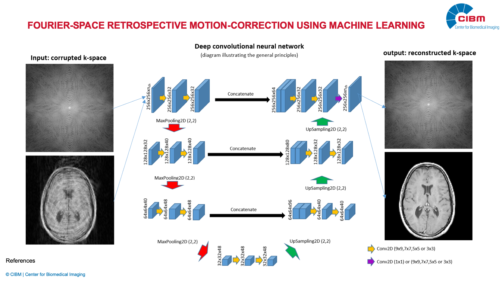
SISMIK: Search In Segmented Motion Input (in) K-space

Description: To tackle the problem of motion artifacts in brain MRI, we propose a deep-learning based approach for rigid-body motion estimation directly in k-space coupled with a model-based reconstruction method to avoid the risk of deep hallucinations. By simulating realistic motion artifacts in motion-free brain MRI scans, we have enabled the generation of large datasets, which is a requirement for training data-hungry deep learning models. SISMIK has the potential to be adapted to a wide range of MRI pulse sequences and k-space trajectories. We hope that the research and clinical communities will benefit from our method..

Investigators: Oscar Dabrowski (UNIGE), Sébastien Courvoisier (HUG), Jean-Luc Falcone (UNIGE), Bastien Chopard (UNIGE), Francois Lazeyras (HUG), Frédéric Grouiller (HUG)

Whole brain high-resolution spectroscopic imaging

Description: A new approach to image brain metabolites of the entire cortical convexity in high resolution was developed. The method combines fast MRSI acquisition, compressed sensing and low-rank modeling for constrained reconstruction. The method allows for high-resolution acquisition of  an entire 2D-slice in 6 min and an whole brain 3D-volume in 20 min.

Investigators: Francois Lazeyras (HUG), Antoine Klauser (UNIGE), Sébastien Courvoisier (UNIGE)

Collaborators: Ovidiu Andronesi (MGH, Harvard Medical School, Boston, MA, USA), Bernhard Strasser (Vienna General Hospital, Vienna, Austria)

High-resolution MRSI by artificial intelligence

Description: Using new artificial intelligence tools to provide a full spectral analysis framework for HR-MRSI applications. The method combines realistic synthetic spectral generation, neural networks models, and credibility estimation. This approach has excellent quantification performance, with a processing time more than 10’000 times faster compared to standard fitting methods.

Investigators: Francois Lazeyras (HUG), Sébastien Courvoisier (UNIGE), Antoine Klauser (UNIGE), Michel Kocher (HEIG-VD)

Collaborators: Peter Lichard (EPFL), Dawood Ahmed (HEIG-VD), Jonathan Aeschimann (HEIG-VD)

Motion robust acquisition for neonatal imaging

Description: High-resolution MRI is prone to motion degradation due to its lengthy acquisition. This limitation is particularly acute in neonatal imaging, where subject’s position cannot be controlled over time. We have implemented two techniques to reduce motion effects: the first one is to split the long-lasting high-resolution image in several mutually orthogonal low-resolution images, and reconstruct the high-resolution image by super-resolution; the other one is to apply FID-navigation method to asses motion and to compensate it a posteriori.

Investigators: Francois Lazeyras (HUG), Michel Kocher (HEIG-VD), Ceren Askin (UNIGE

Collaborators: Petra S Hüppi (HUG), Tobias Kober (Advanced Clinical Imaging, Siemens Healthineers, Lausanne), Simon Warfield (Boston Children’s Hospital, Boston, MA, USA)

White-matter microstructure maturity using diffusion MRI

Description: This clinical research aims at assessing white matter development of premature baby using advanced diffusion MRI. The overall aim is to better understand brain development and changes of structural connectivity associated with very high prematurity.

Investigators: Petra S Hüppi (HUG), Joana Sà De Almeida (UNIGE), Laura Gui-Lévy (UNIGE), Francois Lazeyras (HUG)

Collaborators: Cyril Poupon (Neurospin, CEA Saclay, Paris, France)

Viability diagnostic of marginal graft by MRI and 31P MRS

Description: Marginal organs (after cardiac arrest, elderly donors) are increasingly considered as an alternative to living donors in order to compensate for the chronic shortage of donors. In order to reduce the risk of graft malfunction, we have developed a technology which allows 1) to resuscitate the organ (kidney) using an MRI-compatible perfusion machine, 2) to perform a complete diagnosis which allows to visualize the kidney, to measure the perfusion and to measure the synthesis of adenosine triphosphate (ATP) by 31P MRS. Alternatively, we study the effect of sulfite oxyde (H2S) as protection agent against warm ischemia and storage.

Investigators: Francois Lazeyras (HUG), Alban Longchamp (CHUV), Antoine Klauser (UNIGE), Julien Songeon (UNIGE), Thomas Agius (CHUV), Jean-Marc Corpataux (CHUV)

Collaborators: Léo Bühler (UniFr), Christian Toso (HUG), Cecil H Charles and Brian J Soher (Duke Medical Center, Durham, NC, USA), Deng Shaoping (Sichuan People’s hospital, Chengdu, China)

In vivo oxidative stress assessment by 31P MRS

Description: Oxidative stress of living tissue can be assessed by several means, including ADP/ATP ratio, NADH/NAD+ ratio and intracellular pH via  31P MRS. The major issue in vivo, on clinical MR systems (3T) , is the lack of spectral resolution and the low SNR that prevents resolving overlapping MR signals. The known MR resonance dynamics of these molecular moieties enables us to predict their behavior and to propose adapted fitting models.

Investigators: Francois Lazeyras (HUG), Julien Songeon (UNIGE), Antoine Klauser (UNIGE), Sébastien Courvoisier (UNIGE)

Collaborators: Laurence Marcourt (UNIGE), Jean-Pierre Wolf (Applied Physic Group (GAP), UNIGE)

Fourier-space retrospective motion-correction using machine learning

Description: Using the fundamentals of k-space properties and MRI acquisition schemes, we aim to develop a complete motion artifact correction framework using machine learning. Indeed, knowing the acquisition sequences, we expect to find corrupted or misplaced datapoints, allowing us to improve the reconstructed image quality. A successfully implemented method could dramatically reduce acquisitions rejection and  would be, for example, a major improvement for neonatal MRI.

Investigators: Francois Lazeyras (HUG), Bastien Chopard (UNIGE), Sébastien Courvoisier (HUG), Oscar Dabrowski (UNIGE), Jean-Luc Falcone (Computer science department, UNIGE)