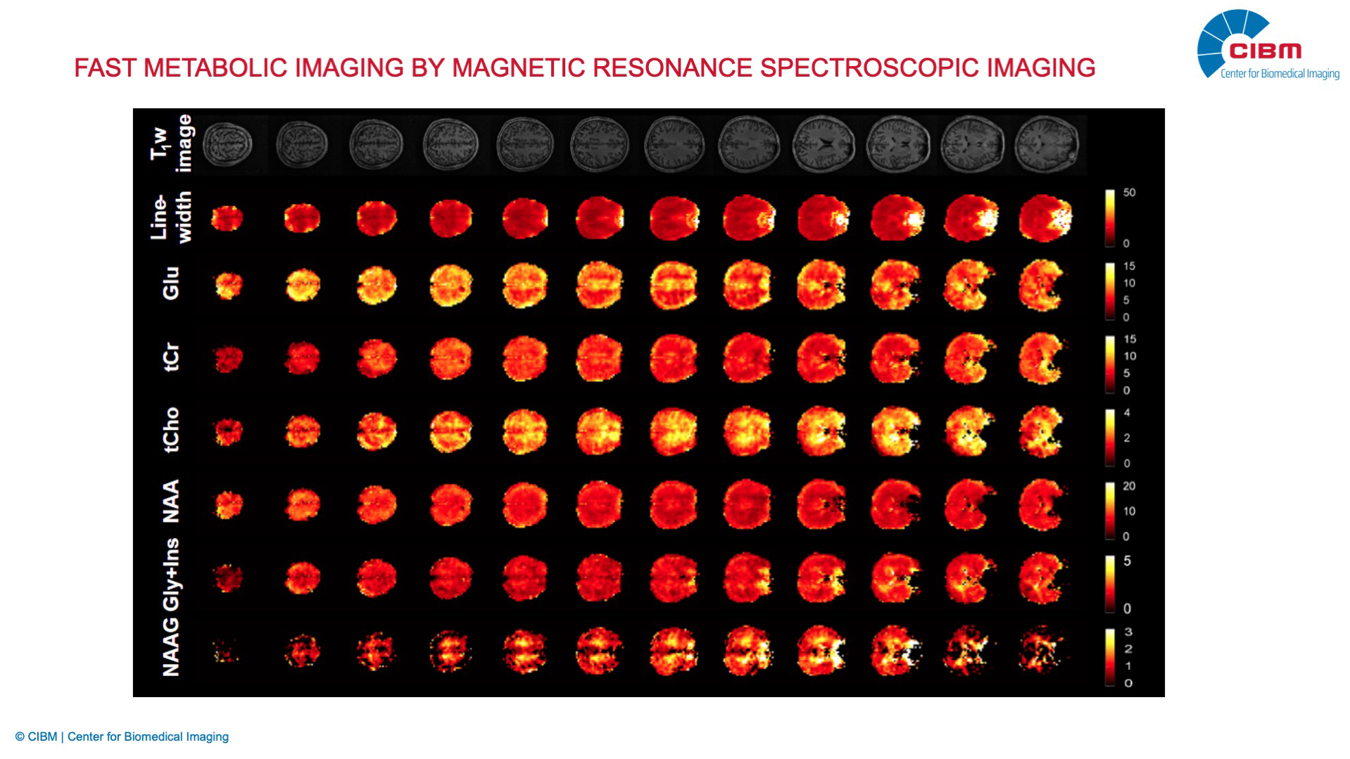
Dynamic 2H MRSI measurement of brain oxidative metabolism
Description:
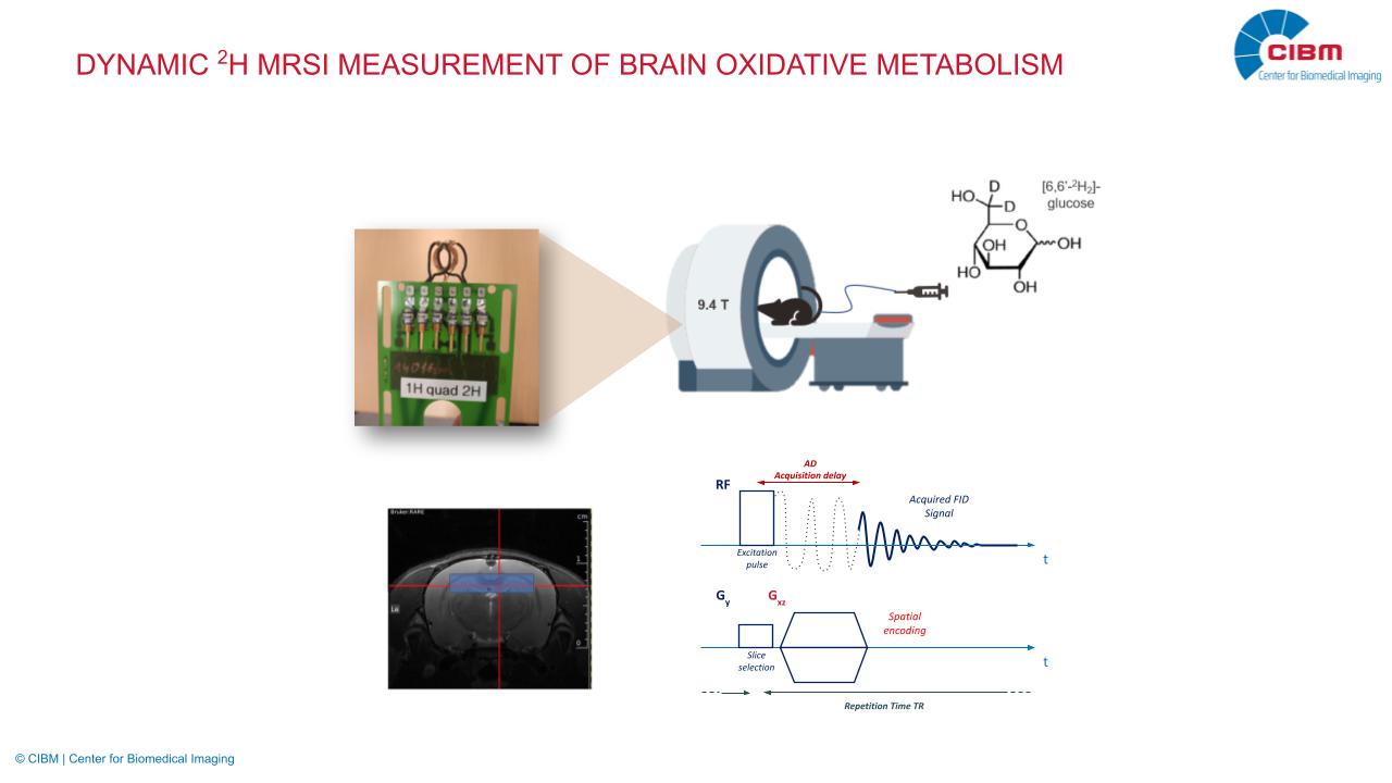
Deuterium imaging (2H-MRS/MRSI) emerged as a promising in vivo tool to study glucose metabolism by the use of deuterium-labelled substrates. Although research to date has shed some light on the usefulness of deuterium imaging in animal models and humans, current 2H-MRSI acquisitions and reconstructions are often limited to static acquisitions at labelling steady-state.
To enable dynamic measurement of deuterium turnover through the brain oxidative metabolic pathway, our research focuses on implementing accelerated MRSI strategies to 3D 2H-MRSI. All developments are aiming at obtaining dynamic and quantitative 3D images of glucose metabolites 2H turnover, enabling further quantitative determination of cerebral oxidative metabolic rate of glucose CMRglc(ox) through advanced metabolic modelling..
Investigator: Bernard Lanz, Alessio Siviglia, Cristina Cudalbu, Gianna Nossa, Thanh Phong Lê (EPFL).
Collaborators: Bernhard Strasser, Fabian Niess, Wolfgang Bogner (Medical University of Vienna)