The new research article, “Partial‐volume modelling reveals reduced gray matter in specific thalamic nuclei early in the time course of psychosis and chronic schizophrenia” Yasser Alemán‐Gómez et al, 10 July 2020, is published by Wiley, https://doi.org/10.1002/hbm.25108 and supported by CIBM Center for Biomedical Imaging.
Dr. Meritxell Bach Cuadra, Head of CIBM SP CHUV-UNIL Section and Medical Image Analysis Laboratory (MIAL), said “I am very proud of this paper and I would like to thank the CIBM”.
She went on to say, “This publication is the result of amazing work by my post doctoral researcher at the time Dr. Yasser Aleman-Gomez in an interdisciplinary collaboration with the CHUV Psychiatry Department that we started with clinical researcher Dr. Philipp Baumann and Post-doctoral researcher Dr. Pascal Steullet with the support of long term CIBM collaborators Prof. Dr. Kim Q. Do & Prof. Philipp Conus from the CHUV Centre de neurosciences psychiatriques (CNP)”.
The research was launched with a grant obtained from the University of Lausanne (UNIL) Faculté de biologie et de médecine (FBM) interdisciplinary projects.
Abstract
The structural complexity of the thalamus, due to its mixed composition of gray and white matter, make it challenging to disjoint and quantify each tissue contribution to the thalamic anatomy. This work promotes the use of partial‐volume‐based over probabilistic‐based tissue segmentation approaches to better capture thalamic gray matter differences between patients at different stages of psychosis (early and chronic) and healthy controls. The study was performed on a cohort of 23 patients with schizophrenia, 41 with early psychosis and 69 age and sex‐matched healthy subjects. Six tissue segmentation approaches were employed to obtain the gray matter concentration/probability images. The statistical tests were applied at three different anatomical scales: whole thalamus, thalamic subregions and voxel‐wise. The results suggest that the partial volume model estimation of gray matter is more sensitive to detect atrophies within the thalamus of patients with psychosis. However all the methods detected gray matter deficit in the pulvinar, particularly in early stages of psychosis. This study demonstrates also that the gray matter decrease varies nonlinearly with age and between nuclei. While a gray matter loss was found in the pulvinar of patients in both stages of psychosis, reduced gray matter in the mediodorsal was only observed in early psychosis subjects. Finally, our analyses point to alterations in a sub‐region comprising the lateral posterior and ventral posterior nuclei. The obtained results reinforce the hypothesis that thalamic gray matter assessment is more reliable when the tissues segmentation method takes into account the partial volume effect.
Access the article here: https://onlinelibrary.wiley.com/doi/full/10.1002/hbm.25108
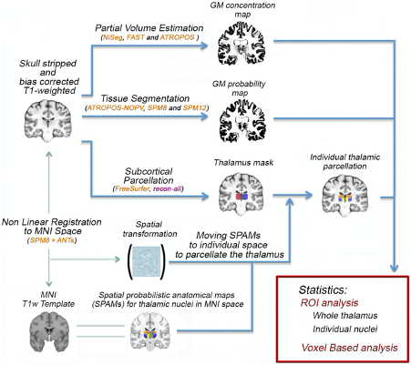
FIGURE 1 Image processing workflow summary. The estimation of gray matter within the thalamus is performed by using six different segmentation methods. The analysis is done at three different anatomical scales: whole thalamus, thalamic regions and voxel‐wise.