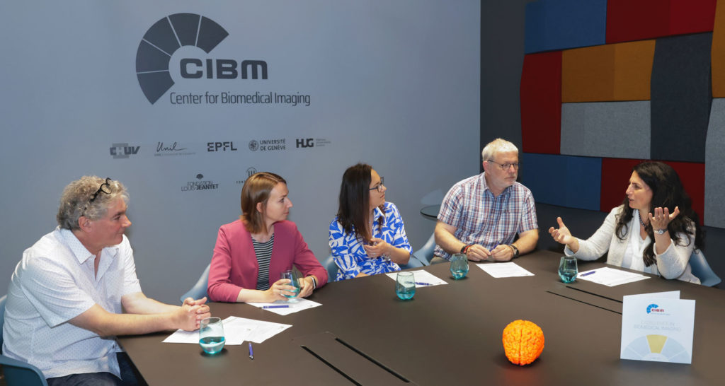
In a remarkable demonstration of the CIBM Center for Biomedical Imaging collaborative spirit and scientific determination, a multi-institutional team of four researchers from CIBM’s founding partner institutions, École polytechnique fédérale de Lausanne (EPFL), University of Lausanne (UNIL), Lausanne University Hospital (CHUV), University of Geneva (UNIGE) and Geneva University Hospitals (HUG), has been awarded a prestigious four-year multi-disciplinary grant from the Swiss National Science Foundation (SNSF) for their ground breaking project titled “Shedding Light on the Invisible: Mapping Brain Microstructure Using Diffusion Magnetic Resonance Spectroscopic Imaging.”
The 3.7 million CHF grant is a hard-earned victory, paving the way for innovative research that could unlock new insights into the intricate workings of the brain.
Leading the project are Dr. Cristina Cudalbu from the CIBM MRI EPFL Animal Imaging and Technology Section, Prof. Ileana Jelescu from CHUV Radiology Department, Prof. Olivier Braissant from CHUV Clinical Chemistry Department, both professors at UNIL Faculty of Biology and Medicine, Prof. Stéphane Sizonenko from UNIGE Faculty of Medicine Department of Pediatrics and Gynecology-Obstetrics and project partner Dr. Julien Valette from the Molecular Imaging Research Center (MIRCen) in France.
What does this grant mean for you?
Cristina Cudalbu: A dream came true. I remember the first discussion I had with Olivier in 2019 about the possibility to quantify the diffusivity of glutamine and its direct link with astrocyte morphology in vivo using the organoids produced by Olivier. Since then some time passed, we managed to create our synergistical team with a multidisciplinary background and now we are ready to start this new adventure. We will be mapping and validating brain microstructural changes via water and metabolite diffusivity in the healthy and diseased developing brain. I feel extremely honored and lucky to work at CIBM in this multidisciplinary environment with these amazing researchers.
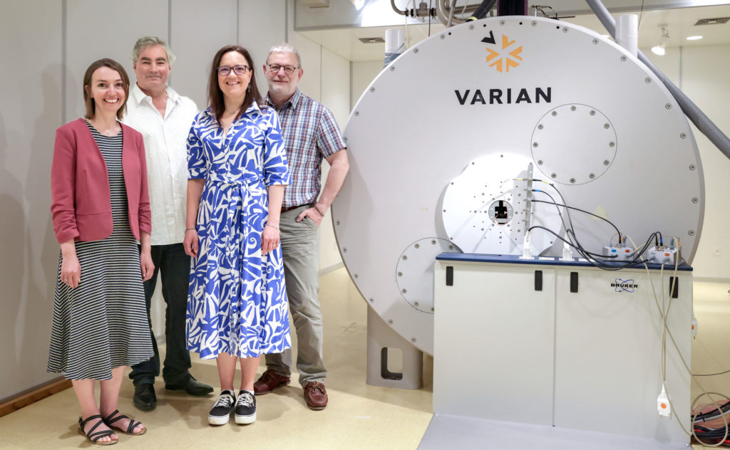
The project’s ambitious goal is to push the boundaries of non-invasive brain microstructure and metabolite quantification by combining novel diffusion MR Spectroscopic Imaging (dMRSI) acquisitions and diffusion modelling techniques. This cutting-edge approach aims to quantify and validate microstructure through the mobility of water and metabolites at cellular and sub-cellular scales across the brain.
To validate their findings, the team will utilise two-photon microscopy and mass spectrometry, applying their methods to 3D organotypic brain cell cultures and animal models. This multifaceted approach underscores the project’s multidisciplinary nature, drawing upon expertise from various institutions and disciplines.
What excites you most about the potential impact of this project?
Ileana Jelescu: I have been using diffusion of water molecules to probe microstructure for over ten years. While we have made significant progress on the level of biophysical modeling and on the microstructure features that we estimate non-invasively, we are intrinsically limited in specificity by the fact that water is everywhere in the tissue. The combination of diffusion of water and diffusion of cell-specific metabolites is particularly exciting in terms of the new information it can reveal about the microstructure. Also excited about the potential to uncover pathological changes using these new techniques.
Olivier Braissant: The very challenging measure of metabolites in single living specific brain cells making use of a 2-photon microscopy approach coupled then to mass spectrometry, which will serve to validate the dMRSI data, will certainly unravel so far unknown aspects of CNS physio(patho)logy.
Why is this work important for improving patient care?
Olivier Braissant: This multi-disciplinary project, developing very complementary cutting-edge technologies to unravel cellular microstructure and metabolism in the normal and pathological brain, will certainly pave the way to more precise diagnostics for the patients, and to the development of new therapeutic approaches.
The project will foster further collaboration among institutions in the Léman region, with CHUV, UNIL, EPFL, UNIGE and HUG working synergistically to develop these cutting-edge methodologies in brain research.
What are the benefits of having researchers from different backgrounds collaborating on this project?
Stéphane Sizonenko: The main benefits are that each partner will bring his specific expertise into the project that will enhance discoveries and advancement of the project, giving strong scientific data that will support the development and validation of this new and cutting-edge imaging methodology at the center of the project. Each of the teams are clearly very complementary with their own neuroscience background and research approach which makes this project unique encompassing a global approach.
Further, it will foster exchange of ideas for advancement of the project itself and will also be a major source of education and ideas for the younger researchers that will be implicated in the project.
This multidisciplinary endeavour represents a significant milestone, highlighting the collaborative spirit and determination that drive scientific progress forward, even in the face of initial setbacks.