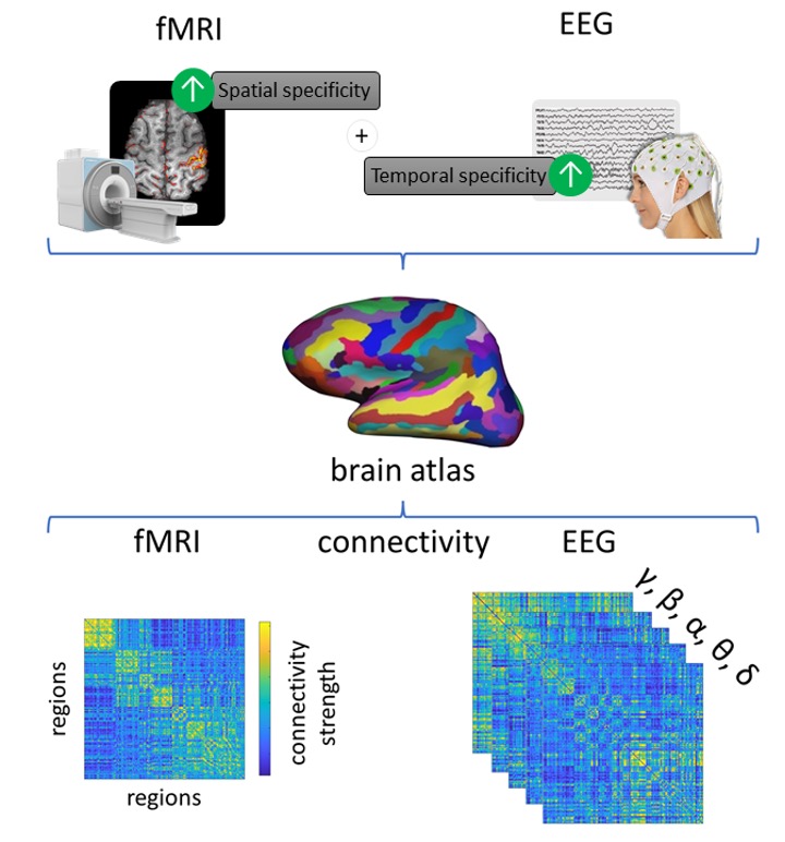
Magnetic resonance imaging (MRI) has become an indispensable tool for medical imaging. Functional MRI (fMRI) measures the neurons’ energy consumption of the brain. As this is an indirect measure of brain activity the exact relationship with neuronal activity needs to be clarified, notably for clinical applications. The electroencephalogram (EEG) captures electrical activity of the brain and is routinely used in clinical practice, notably for the diagnosis of epilepsy. EEG has higher temporal precision but lower special accuracy than MRI and new approaches are needed to combine information of both modalities.

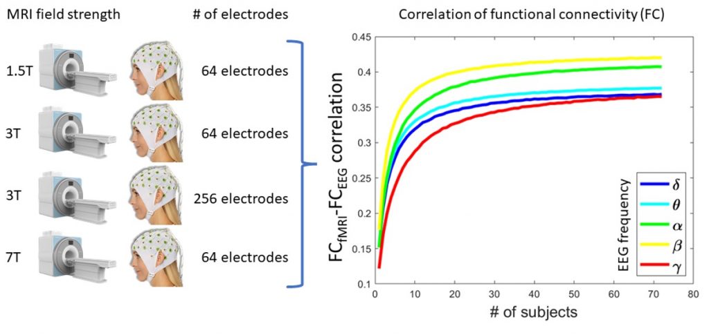
With the goal of better understanding the link between EEG and fMRI, researchers from the CIBM Center for Biomedical Imaging at HUG, EPFL and the University of Geneva have studied the reliability of measuring brain networks derived from both EEG and fMRI (the so called functional connectivity networks, see illustration). In a multicentric approach, the researchers compared data acquired with the 3 Tesla (HUG-UNIGE) and 7 Tesla (EPFL) MRI platforms of CIBM, as well as two datasets available from international brain imaging centers (Paris, London). This allowed for the most comprehensive multi-field and multi-system study to date on EEG-fMRI.

The researchers observed a reliable functional connectivity between EEG and fMRI networks for all datasets, depending on the number of subjects used to estimate the brain network. This result validates the close link between EEG and fMRI. Especially high-field MR data acquired at 7T holds the promise to push forward the spatial precision of fMRI providing unprecedented detailed insights into brain function in healthy subjects and patients. In combination with EEG, routinely used in clinical day-to-day care, this approach is one step forward to using combined recordings in a clinical setting, enabling higher precision to develop new approaches for personalized medicine.
The authors of this international and cross institutional research collaboration are as follows:
Jonathan Wirsich a , ∗ , João Jorge b , c , Giannina Rita Iannotti a , Elhum A Shamshiri a, Frédéric Grouiller d , Rodolfo Abreu e , f , François Lazeyras g , Anne-Lise Giraud h , Rolf Gruetter b , g , i , Sepideh Sadaghiani j , k , Serge Vulliémoz a
a EEG and Epilepsy Unit, University Hospitals and Faculty of Medicine of Geneva, University of Geneva, Geneva, Switzerland
b Laboratory for Functional and Metabolic Imaging, École Polytechnique Fédérale de Lausanne, Lausanne, Switzerland
c Systems Division, Swiss Center for Electronics and Microtechnology (CSEM), Neuchâtel, Switzerland
d Swiss Center for Affective Sciences, University of Geneva, Geneva, Switzerland
e ISR-Lisboa/LARSyS and Department of Bioengineering, Instituto Superior Técnico –Universidade de Lisboa, Lisbon, Portugal
f Coimbra Institute for Biomedical Imaging and Translational Research (CIBIT), Institute for Nuclear Sciences Applied to Health (ICNAS), University of Coimbra, Coimbra, Portugal
g Department of Radiology and Medical Informatics, University of Geneva, Geneva, Switzerland
h Department of Neuroscience, University of Geneva, Geneva, Switzerland
i Department of Radiology, University of Lausanne, Lausanne, Switzerland
j Beckman Institute, University of Illinois at Urbana-Champaign, Urbana, IL, United States
k Psychology Department, University of Illinois at Urbana-Champaign, Urbana, IL, United States
The original article (Wirsich et al. 2021, NeuroImage) can be accessed here:
https://www.sciencedirect.com/science/article/pii/S105381192100141
