Eye movements are a major obstacle for imaging the eye, especially in the cases of children, the elderly or patients with eye disease.
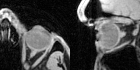
Supported by the Swiss National Science Foundation, a team of researchers composed of neuroscientists, engineers, mathematicians, and optometrists from the Radiology Department of the Lausanne University Hospital Center (CHUV) and University of Lausanne (UNIL), as well as, Fondation Asile des Aveugles (FAA), CIBM EEG and MRI CHUV-UNIL Sections, conducted a study published in Elsevier’s journal Progress in Neurobiology which shows that it is possible to take magnetic resonance imaging pictures of the eye as it moves, revealing for the first time simultaneous details about the eye, its musculature, and oculomotor properties.
“This is the first-time scientists are able to image the eye while it moves, establishing a link between behaviour and anatomy. This will open a new field of ophthalmic MRI, where we will be finally able to combine multiple assessments within a single, fast, session. The applications are infinite. They span from medical diagnosis to eye-brain mechanisms,” explains Dr. Benedetta Franceschielo, a post-doctoral fellow and lead author of the study, who works in the team of Professor Micah Murray, principal investigator of this project.
This innovative technique is based on recent advances in magnetic resonance imaging that allow for taking many snapshots of an object that repeatedly moves, such as the beating heart or moving eye. Professor Stuber emphasizes, “Five years ago, this research would not have been feasible. Only at the confluence of staggering progress in hardware, software, and methodology development in a university setting did this become possible.”
Professor Murray emphasizes the wider impact of this new technological advancement: “The necessity to fixate ordinarily restricts the range of compliant participants; something often challenging for paediatric and aged populations alike. We have removed the onus of maintaining central fixation. Because we used standard MRI equipment and eye-tracking hardware, the approach we have developed and validated is readily deployed to the broad scientific community and impacts not only the breadth of participant inclusion, but also the extent of naturalistic paradigms that can be investigated. We can now study how perceptions are constructed as we move our eyes across a scene, an artwork, or as we read. A further advantage of our method is that eye position can be followed even when the eyes are closed. This would open new possibilities in fields such as sleep and dream research as well as more generally for understanding brain activity in disorders of consciousness.”
The team is now refining this technique to optimise it and make it versatile for clinical application in the field of ophthalmology. This invention not only represents a turning point in the way we study the eyes, but also in cognitive neuroscience. It has also recently demonstrated its value by allowing the creation of new protocols both in the field of diagnosis in ophthalmology as well as in visual rehabilitation.
Reference: Franceschiello B*, Di Sopra L*, Minier A, Ionta S, Zeugin D, Notter M P, Bastianseen J A M, Jorge J, Yerly J, Stuber M, Murray M M (2020). 3-Dimensional Magnetic Resonance Imaging of the Freely Moving Human Eye. Progress in Neurobiology. https://doi.org/10.1016/j.pneurobio.2020.101885