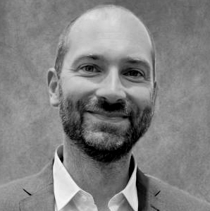
Visitors Talk: Thomas Christen, Grenoble Institute for Neurosciences
Hosted by Jean-Noël Hyacinthe, Scientist at the Laboratory of Functional and Metabolic Imaging, EPFL, we are pleased to invite you to attend the CIBM Visitors Talk on November 13th at 11:00 CET by Thomas Christen from the Grenoble Institute for Neurosciences who will be sharing on “Estimating 3D microvascular structures and blood oxygenation with MR Fingerprinting”.
Date and time: Friday, November 13th, 2026 – 11:00 CET
Dr Thomas Christen is an MR physicist, deputy director of the neuroimaging research team at Grenoble Institute for Neurosciences (GIN), and director of the joint research and service IRMAGE facility located in the GIN and the Grenoble University Hospital Center. He earned his Ph.D. in applied physics from Grenoble University, followed by postdoctoral training and research position at Stanford University. His research focuses on developing new MRI techniques to better understand human brain function and diagnose brain diseases. This includes improved acquisition schemes (MRI sequences, hardware) and post-processing algorithms for studying stroke, cancer and traumatic brain injuries.
TALK: Estimating 3D microvascular structures and blood oxygenation with MR Fingerprinting
Abstract
The Magnetic Resonance Fingerprinting (MRF) framework allows measurements of several tissue properties simultaneously with a single fast and efficient MR sequence combined with precomputed MR signal simulations (dictionary approach). Despite the initial focus on relaxometry estimates, the method is not limited to a single excitation pattern nor the measurement of the transverse or longitudinal relaxation times only. It is theoretically possible to quantify any tissue property, as long as the MRF sequence is sensitive to the parameters of interest, provides different fingerprints for different tissue properties, and that the MRF simulations are realistic enough to capture the signal variations.
Over the last decade, we have explored the potential of MRF to quantify brain microvascular structures and function by combining Blood Oxygen Level Dependent (BOLD)- sensitive sequences with biophysical simulations based on ex vivo 3D high-resolution microscopy data. In this talk, we will present an overview of key vascular MRF studies, emphasizing advancements in geometrical models for vascular simulations, novel contrast-free sequences, and advanced reconstruction techniques. We will discuss both preclinical and clinical applications, as well as the possibility to extend the analysis to standard brain MR exams.
Seminar Chair
Jean-Noël Hyacinthe
Scientist, Laboratory of Functional and Metabolic Imaging