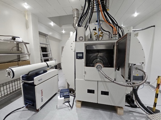
The CIBM MRI EPFL Section successfully enhanced and extended its 9.4T preclinical horizontal-bore MRI with cutting-edge MR electronics, gradients, radiofrequency coils and a PET insert. This marks a significant step towards ultra-high field MR-PET imaging for 3D mapping of function, structure and metabolism; a unique setup in Switzerland.
Magnetic resonance imaging and spectroscopy (MRI, MRS) together with Positron Emission Tomography (PET) have made tremendous advancements as a diagnostic modality. The new equipment enhances the state-of-the-art infrastructure of the CIBM MRI EPFL Animal Imaging and Technology Section to continue leading the way in pre-clinical biomedical imaging research.
Key elements of the upgrade package include multichannel radio frequency transmission and reception capabilities, stronger imaging and shimming gradients, a cryogenic radiofrequency coil providing an unmatched boost in sensitivity, and a PET insert. This setup allows for concomitant in vivo MR-PET examinations for simultaneous, complementary and multi-contrast information on organ structure, function and metabolism; a setup unique in Switzerland.
The CIBM pre-clinical imaging researchers rapidly transitioned to the new equipment, incorporating it into their routine studies to acquire a wide range of anatomical, metabolic and microstructural information in vivo.
This new-generation ultra-high-field MR-PET scanner will significantly strengthen the scientific landscape in the region and all over Switzerland and Europe. It is expected that the broader scientific community will benefit from the new research developed using the new MR-PET scanner.
CIBM extends its gratitude to all scientific collaborators who supported this project by highlighting the potential benefits of the new equipment for their research endeavours. Special thanks are due to EPFL for their co-funding of the equipment and their invaluable assistance with the renovations and adaptations needed to accommodate this cutting-edge system in its current location.

Upgraded 9.4T MR-PET system

Anatomical (T2 weighted MRI) and microstructural (diffusion tensor imaging) measurements of the rat brain using the cryogenic radiofrequency coil

MR spectroscopic imaging maps and corresponding spectra acquired with the cryogenic radiofrequency coil

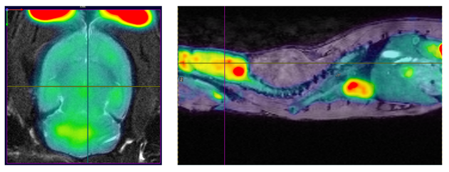
First dynamic MR-PET examinations in the rat brain and body using 18F-FDG

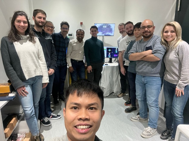
CIBM MRI EPFL pre-clinical imaging researchers and Bruker team after successful MR-PET training
The headquarters of the CIBM Center for Biomedical Imaging at EPFL is home to the latest high-end multimodal bioimaging infrastructure (ultra-high field magnetic resonance imaging systems (9.4T and 14.1T), electron paramagnetic resonance (EPR) spectroscopy, etc.), nurtures collaborations and provides expertise to academic institutions, startups and pharmaceutical companies from all over Switzerland (EPFL, UNIL, CHUV, UNIGE, HUG, ETHZ, UBern, UBasel) and Europe. Access to highest level and well-maintained facilities for data acquisition and processing is fundamental for researchers to achieve timely progress and go beyond the current state-of-the-art worldwide which is aligned with CIBM’s strategic roadmap.
