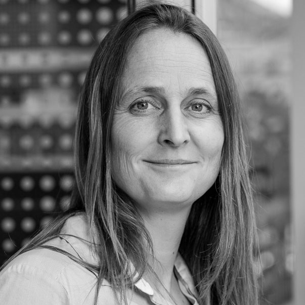
On February 21 2024, Wietske van der Zwaag, from the Spinoza Centre for Neuroimaging, Amsterdam, The Netherlands, shared her talk on “High resolution 7T to image the human cerebellum” at the Campus Biotech in Geneva.
Insights
Ultra-high field (7T) MRI offers both higher signal-to-noise ratios and higher sensitivity to the functional BOLD signal used in fMRI. These can be traded in for smaller voxel sizes and this makes 7T MRI exquisitely suited for high-resolution (functional) imaging.
The human cerebellum is an intricately shaped brain area at the back of the head, which plays a role in all major brain networks. It is involved in a wide range of functions including visual control, emotion and language, but remains understudied, in part because its cortex cannot be resolved with standard MRI techniques.
In this talk, she discussed the benefits of 7T, and specifically the higher spatial resolution, can be beneficial for imaging the human cerebellum in-vivo.

Wietske van der Zwaag
Spinoza Centre for Neuroimaging, Amsterdam, The Netherlands
I joined the Spinoza Centre for Neuroimaging in 2015. In 2019, I formed my own group here, working at the boundary between MR-development and neuroscicence. Previously, I worked with 7T-MRI at the École Polytechnique Fédérale de Lausanne (EPFL) in Switzerland. I did my PhD at the Sir Peter Mansfield Imaging Centre (SPMIC) in Nottingham in the United Kingdom under supervision of Richard Bowtell and Sue Francis .
Our research is centred on best harnessing the strong points of 7T in neuroimaging. I am especially interested in functional MRI of finely organized brain structures, such as the human cerebellum. The use of ultra-high field scanners (7T) allows the study of the human brain at unprecedented resolution and is ideal for studying this intricately structured brain region. In our work, we look for solutions for the technical problems that also form a part of scanning at 7T, to best profit from the technical advantages offered by 7T MRI.
