
CIBM Annual Symposium 2026
The CIBM Annual Symposium 2026 will take place on 2 December 2026 for the first time at CHUV in the Cesar Roux auditorium. The annual event brings together members of the biomedical imaging community in Switzerland and beyond allowing them to exchange during an exciting and intellectually stimulating day filled with insightful talks and poster presentations.
PROGRAM
Registration opens at 8h30
9:00 Welcome Breakfast: Posters and Demos
10:00 Welcome & Opening
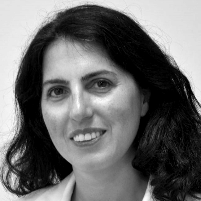
Pina Marziliano
CIBM Executive Director
MORNING SESSIONS – Chaired by TBC
10:15 – The Future of Radiology
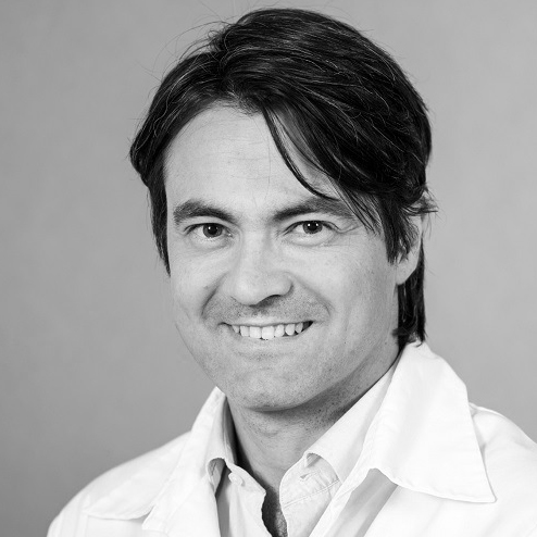
Paolo Machi
HUG – UNIGE
ABSTRACT
The future of radiology may be shaped as much by organizational transformation as by technological innovation. As artificial intelligence evolves beyond isolated image-analysis tools, coordinated networks of specialized AI agents could support the entire patient journey, connecting diagnosis, clinical decision-making, image-guided intervention, and longitudinal follow-up. Drawing on diagnostic and interventional radiology, this presentation explores a framework for AI-orchestrated radiology. Central to this vision is the Patient Orchestrator: a coordination layer that preserves longitudinal clinical context and connects specialized AI capabilities according to each patient’s evolving needs. These capabilities extend beyond imaging to procedural assistance, departmental operations, research, education, and quality assurance. Such integration could reduce information fragmentation, strengthen multidisciplinary collaboration, and enable departments to become learning environments in which routine clinical activity continuously informs improvements in care. Realizing this vision will require interoperable infrastructure, prospective validation of interacting AI systems, transparent governance, and sustained physician oversight. Distinguishing current capabilities from emerging concepts, this presentation outlines a progressive pathway toward more connected, adaptive, and patient-centered radiology. The ultimate ambition is to give clinicians greater capacity for clinical reasoning, innovation, and compassionate care.
BIO
Paolo Machi, MD, PhD, is Head of Interventional Neuroradiology at Geneva University Hospitals, Associate Professor at the University of Geneva, and Professor of Radiology at Mayo Clinic. Since December 1, 2026, he has served as Academic Director of the Department of Radiology and Medical Informatics at the University of Geneva. He is the founder and director of the Brain Endovascular Therapeutics R&D Lab at Campus Biotech, Geneva. With a PhD in mechanical engineering, he combines clinical expertise with translational research in artificial intelligence, robotics, and medical device development. His research explores AI-supported decision-making, robotic interventions, and patient-specific simulation to improve neurovascular care. His work bridges medicine, engineering, and healthcare innovation, with a focus on shaping the future of diagnostic and interventional radiology.
10:45 – Intraoperative gamma imaging, a new tool for the complete radio-guided surgery workflow: 3D localisation, completeness check and ex-vivo verification
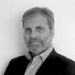
Domenico Della Volpe
UNIGE
ABSTRACT
Image-guided surgery includes a wide range of imaging modalities, each with its own strengths and limitations. In soft-tissue surgery, standard augmented reality (AR) relies on virtual 3D models built from preoperative images (CT, MRI, ultrasound), but rigid registration fails when tissues move and deform. Optical modalities, such as fluorescence or spectral imaging, see only a few millimetres below the surface. Nuclear imaging offers unmatched specificity in guiding the surgeon to radiotracer-avid targets. However, SPECT and PET need bulky equipment and heavy logistics, while portable gamma probes give only acoustic guidance, with no image and no depth.
Smart multimodal approaches can overcome the limits of each single modality. This talk presents a new concept: a wireless, handheld gamma camera that reconstructs the tracer distribution in 3D from free-hand scans and shows it directly on the patient through AR. POSICS is developed at the University of Geneva in collaboration with the PINLab at HUG, with HEPIA and Campus Biotech contributing to the 3D and AR part. The same device follows the whole procedure: before resection, to locate the target in 3D; during surgery, with the image overlaid on the patient; before closing, to check that the target has been completely removed; and after excision, to verify the specimen ex vivo. I will show first 3D results in mice.
BIO
Domenico della Volpe is Associate Professor of Experimental Particle Physics at the University of Geneva. He holds an M.Sc. (magna cum laude, 1994) and a Ph.D. (1998) in Physics from the University of Naples Federico II.
He spent fifteen years on the ATLAS experiment at CERN (1998–2013), working on the design, construction, commissioning, operation and upgrade of the detector and its trigger systems. He was Level-1 Trigger Coordinator, Barrel Muon Trigger Coordinator and Run Manager, and main editor of the ATLAS Phase-II Upgrade Letter of Intent. He still sits on the ATLAS Upgrade Project Management Office review panel. As an ATLAS author, he shares the 2025 Breakthrough Prize in Fundamental Physics.
He then moved to gamma-ray astronomy. He was Lead System Engineer of the CTAO Large-Sized Telescope and Project Manager of the SST-1M telescope. Today he coordinates the Swiss CTAO consortium and is Swiss delegate to the CTAO-ERIC Council.
His expertise covers radiation detectors, silicon photomultipliers, gamma-ray imaging, trigger and data-acquisition systems, and systems engineering of large instruments.
He now applies this experience to medicine as co-founder of POSICS, a UNIGE–FBK spin-off project developing a handheld, wireless gamma camera with 3D reconstruction and augmented reality for radioguided cancer surgery, in collaboration with HUG. He is an inventor on its European patent and a Technology Expert for the EAES Image-Guided Surgery committee.
He has co-authored about 1,800 publications, with nearly 320,000 citations and an h-index of 232 (Google Scholar).
11:15 – Combining Imaging and Genetics in Dementia Research
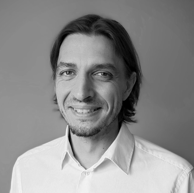
Andre Altmann
UCL Hawkes Institut
ABSTRACT
In this presentation I will present ongoing research that combines genetic, omics and imaging data at different levels to better understand dementia and to achieve more accurate diagnosis and prognosis. The first part will highlight work to understand the genetics of disease progression in dementia; and the second part will focus on integrating genetic data into normative models for better diagnosis and prognosis.
BIO
Andre Altmann is Professor for Biomedical Data Science and AI at UCL’s Department for Medical Physics and Biomedical Engineering and the UCL Hawkes Institute. Following his PhD in Computational Biology at the Max Planck Institute for Informatics in Saarbrücken (Germany), and two post-doctoral positions at the Max Planck Institute of Psychiatry in Munich (Germany) and Stanford University (USA), he started his own lab at UCL in 2015. His research focuses on understanding how molecular biology shapes brain function in healthy and diseased brains and how different modalities can be combined to develop disease biomarkers that assist in early detection and disease progression modeling.
11:45 – Building predictive models for precision oncology across scales, modalities and perturbations
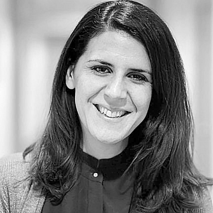
Marianna Rapsomaniki
CHUV – UNIL
ABSTRACT
Recent advances in single-cell and spatial omics, digital pathology, and large-scale perturbation technologies are transforming our ability to characterize cancer across molecular, cellular and tissue scales. Yet translating these heterogeneous data into predictive models that can explain disease mechanisms and anticipate therapeutic response remains a central challenge. In this talk, I will present our group’s vision for building AI models that move beyond descriptive analysis towards virtual representations of cancer capable of integrating diverse data modalities, uncovering biologically meaningful mechanisms, and predicting responses to therapeutic interventions. I will discuss recent work on multimodal representation learning for integrating spatial omics and histopathology, explainable AI for identifying biological and spatial determinants of tumour behaviour, and generative and optimal transport-based models for simulating cellular responses to genetic, drug and immunotherapy perturbations. Together, these approaches aim to bridge biological organization from cells to tissues and patients, enabling a new generation of predictive models that support virtual experimentation, accelerate hypothesis generation, and ultimately advance precision oncology.
BIO
Marianna Rapsomaniki is a tenure-track Assistant Professor at the Biomedical Data Science Center of the Lausanne University Hospital (CHUV) and the University of Lausanne (UNIL) since March 2024, leading the AI for Biomedicine research group. Her research focuses on developing AI and machine learning methods tailored to complex, multimodal biomedical datasets, with the goal of elucidating cancer mechanisms and tumor heterogeneity, from the intracellular to the patient level. She holds a Diploma in Computer Science and a Master’s and PhD in Computational Biology from the University of Patras, Greece, while also being a visiting scholar at ETH Zurich. Prior to joining CHUV/UNIL, she was a Staff Research Scientist and Group Leader at IBM Research Zurich.
12:15 Lunch Break: Posters and Demos
AFTERNOON SESSIONS – Chaired by TBC
14:30 – Trustworthy AI for MRI reconstruction

Michael Unser
CIBM SP EPFL
ABSTRACT
TBC
BIO
Michael Unser is Full Professor with EPFL’s School of Engineering and the academic director of EPFL’s Center for Imaging, Lausanne, Switzerland. His primary areas of investigation are biomedical imaging and applied functional analysis. He is internationally recognized for his research contributions to sampling theory, wavelets, the use of splines for image processing, stochastic processes, and computational bioimaging. He has published over 400 journal papers on those topics. He is the author with P. Tafti of the book “An introduction to sparse stochastic processes”, Cambridge University Press 2014.
He was born in Zug, Switzerland, on April 9, 1958. He received the M.S. (summa cum laude) and Ph.D. degrees in Electrical Engineering in 1981 and 1984, respectively, from the Ecole Polytechnique de Lausanne (EPFL), Switzerland. From 1985 to 1997, he was with the Biomedical Engineering and Instrumentation Program, National Institutes of Health, Bethesda USA, conducting research on bioimaging.
Dr. Unser has served on the editorial board of most of the primary journals in his field including the IEEE Transactions on Medical Imaging (associate Editor-in-Chief 2003-2005), IEEE Trans. Image Processing, Proc. of IEEE, and SIAM J. of Imaging Sciences. He was general co-chair (with Z.P. Liang) for the first IEEE International Symposium on Biomedical Imaging (ISBI’2002), which was held in Washington, DC, July 7-10, 2002. He is also the founding chair of the technical committee on Bio Imaging and Signal Processing (BISP) of the IEEE Signal Processing Society.
Michael Unser received the Dommer prize for excellence in 1981 (1st rank among all EPFL graduates) and the research prize of the Brown-Boveri Corporation (Switzerland) for his Ph.D. thesis in 1984. He is a fellow of the IEEE (1999), an EURASIP fellow (2009), and a member of the Swiss Academy of Engineering Sciences. He is the recipient of several international prizes including five IEEE-SPS Best Paper Awards, two Technical Achievement Awards from the IEEE (2008 SPS and EMBS 2010), the Technical Achievement Award from EURASIP (2018), a Career Achievement Award (IEEE EMBS 2020) and recent the 2025 Norbert Wiener Society Award for his contributions and leadership to the theory and practice of signal processing and biomedical imaging from the IEEE Signal Processing Society. He was awarded three ERC AdG grants: FUNSP (2011-2016), GlobalBioIm (2016-2021), and FunLearn (2021-2026).
Since its creation in 2004, Michael Unser has played a leading role in the CIBM Signal Processing Module and he is the Head of the CIBM SP EPFL Mathematical Imaging Section.
15:00 – Title TBC
15:30 – Title TBC
16:00 – Title TBC
16:30 – BBC: Brain, Body and consciousness
Juliane Britz
University of Fribourg
ABSTRACT
Conscious awareness varies with trial-by-trial variations in spontaneous brain activity and with cyclic fluctuations of bodily signals such as cardiac and respiratory activity. I will present a series of studies in which we investigate brain-body interactions for conscious awareness and will show how 1) baroreceptor fluctuations across the cardiac and respiratory phase determine both the earliest electrophysiological marker and the trajectory of brain activity of awareness, 2) the mode of breathing modulates awareness-related brain potentials and 3) the momentary state of the brain and the body contribute independently to awareness.
BIO
During my postdoc at the CIBM with Christoph Michel, I became interested in the idea of state-dependency in the brain and investigated how the state of the brain determines conscious awareness and in more general notions of the organization of resting-state brain activity. I am now a lecturer at the University of Fribourg and became fascinated by the role of bodily signals on brain activity and have incorporated the role of bodily signals into my two lines of research.
17:00 Closing remarks
17:00 Apéro & Networking
- 02 Dec 2026
- 9:00 am - 6:00 pm
- Cesar Roux Auditorium, CHUV Lausanne